. 2010 Apr;36(4):809-16.
doi: 10.3892/ijo_00000557.

Radiation-inducible silencing of uPA and uPAR in vitro and in vivo in meningioma

Affiliations

Radiation-inducible silencing of uPA and uPAR in vitro and in vivo in meningioma

Venkateswara Rao Gogineni et al. Int J Oncol. 2010 Apr.

Abstract

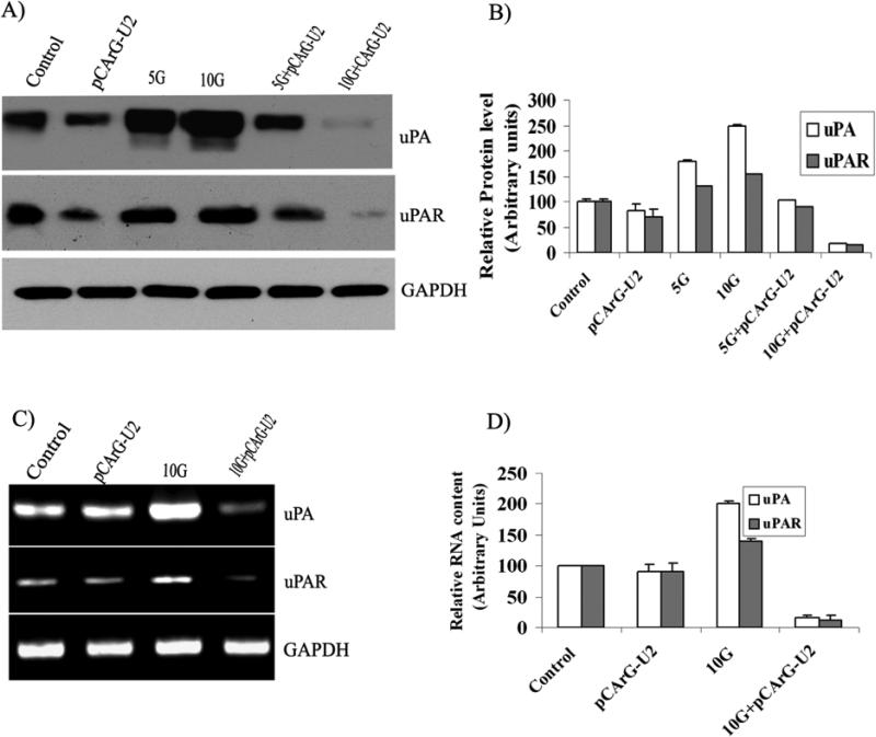
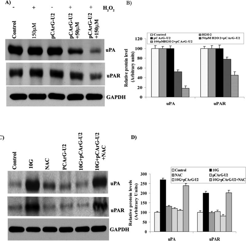
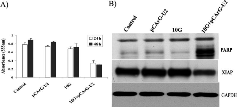
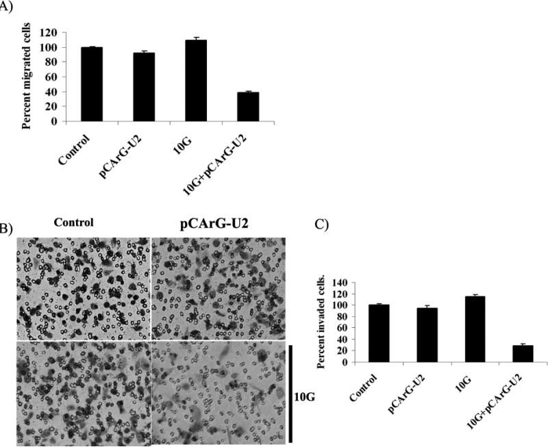
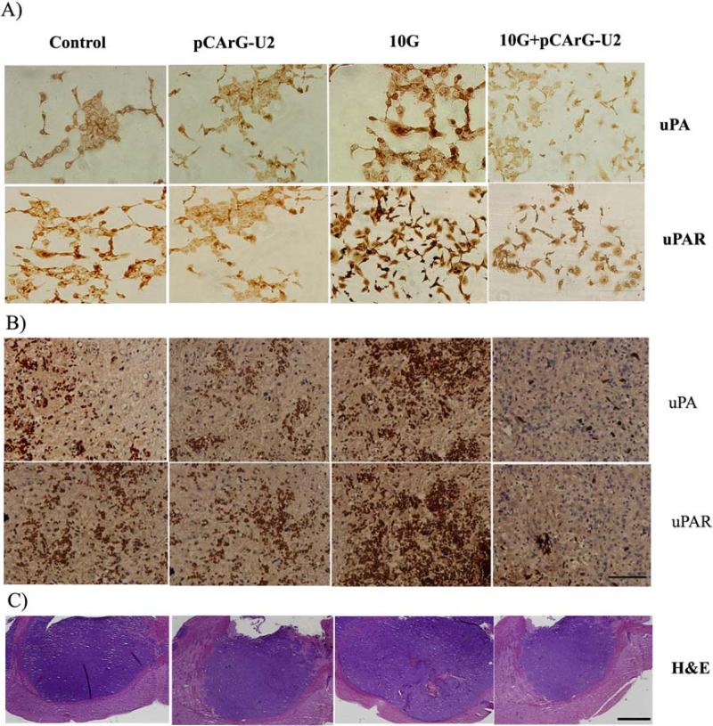
Stereospecific radiation treatment offers a distinct opportunity for temporal and spatial regulation of gene expression at tumor sites by means of inducible promoters. To this end, a plasmid, pCArG-U2, was constructed by incorporating nine CArG elements (in tandem) of EGR1 gene upstream to uPA and uPAR siRNA oligonucleotides in a pCi-neo vector. Radiation-induced siRNA expression was detected in a meningioma cell line (IOMM-Lee). Immunoblotting and RT-PCR analyses confirmed downregulation of uPA and uPAR. A similar effect was observed in transfected cells followed by H2O2 treatment. Moreover, pre-treatment of transfected cells with N-acetyl L-cysteine blocked the silencing of uPA and uPAR, which further confirmed the oxidative damage-mediated downregulation. Cell proliferation assays and Western blot analysis for apoptotic molecules confirmed cell death in a radiation-inducible fashion. Migration and matrigel invasion assays also revealed a marked decrease in migration and invasion. Immunocytochemistry showed a marked decrease in uPA and uPAR levels in transfected and irradiated cells. H&E staining revealed a decrease in the pre-established tumor volume among the animals treated with pCArG-U2 and radiation. Immunohistochemistry of the brain sections established with intracranial tumors also revealed a marked decrease in uPA and uPAR in a radiation-inducible fashion. Taken together, our data suggest pCArG-U2 as a suitable candidate for radiation-inducible gene therapy.


Figures

Figure 1
Figure 1. uPA and uPAR are knocked down in a radiation-inducible manner
(A) IOMM Lee cells were transfected with pCArG-U2 and 24 h later, treated with 5 and 10 Gy. Non-transfected cells were maintained as controls. Cell lysates were collected and equal amounts of proteins were subjected to immunoblotting with uPA and uPAR antibodies. (B) Densitometric analysis of uPA and uPAR western blots. (C) IOMM Lee cells were transfected with pCArG-U2 and irradiated with 10 Gy. 48 h after transfection, total RNA was extracted from the cells as per standard protocols. Reverse transcription PCR analysis was performed using primers specific for uPA, uPAR and GAPDH. (D) Densitometric analysis of relative transcript levels of uPA and uPAR. Results from three independent experiments are shown as means ± S.D (p<0.05).
Figure 2
Figure 2. The inducibility of pCArG-U2 is mediated by oxidative damage
(A) IOMM Lee cells were transfected with pCArG-U2, and after 24 h, treated with different concentrations of H2O2 for another 24 h. Cell lysates were subjected to western blot analysis using uPA and uPAR antibodies. (B) Quantification of uPA and uPAR levels in various treatment groups from the western blots. (C) IOMM Lee cells were transfected with pCArG-U2 and treated with 30 mM N-Acetyl L-Cysteine for 45 min before radiation treatment with 10 Gy. 24 h after radiation treatment, cell lysates were subjected to western blotting with uPA and uPAR antibodies. (D) Densitometric analysis of western blots. Results from three independent experiments are shown as means ± S.D (p<0.05).
Figure 3
Figure 3. Transfection with pCArG-U2 induces apoptosis in a radiation-inducible manner
(A) IOMM-Lee cells were transfected with pCArG-U2 and irradiated with 10 Gy after 12 h. Six hours later, cells were trypsinized, counted and seeded at 5×10³ cells per well in 96-well plates (8 wells per treatment group). After the indicated periods of incubation, MTT assay was performed as described in Materials and Methods, and absorbance values were plotted against the time periods. Data shown are mean ± SD from three different experiments (p<0.05). (B) Cell lysates from pCArG-U2-transfected, irradiated and control cells were used for immunoblot analysis for PARP cleavage and XIAP expression levels using specific antibodies.
Figure 4
Figure 4. Knockdown of uPA and uPAR reduces migration and invasion in a radiation-inducible manner
(A) IOMM-Lee cells were transfected with pCArG-U2 and irradiated after 24 h. Untreated cells were also maintained to serve as the control. Immediately after radiation treatment, 5×10⁴cells were transferred to the upper chamber of 8 μM transwell insert and maintained for another 24 h in serum-free media with complete medium in the lower chamber. Cell migration was computed as the relative number of treated cells migrated compared to the respective controls groups after staining. Values are mean ± SD from three different experiments (p<0.05). (B) IOMM-Lee cells were transfected with pCArG-U2 and irradiated at 10 Gy 24 h after transfection. Untreated cells (mock) were also maintained to serve as the control. After irradiation, the cells were trypsinized and 1×10⁵ cells from each treatment group and controls were cultured in the upper chamber of a transwell insert coated with Matrigel (1 mg/mL). Representative pictures were taken after 24 h of incubation. (C) The number of cells in three different fields for each sample was counted. The percent invasion of cells transfected with pCArG-U2 was analyzed and compared with the untreated (mock) cells. Values are mean ± SD from three different experiments (p<0.05).
Figure 5
Figure 5. Immunocytochemistry and immunohistochemistry reveal decreased uPA and uPAR
(A) IOMM Lee cells were seeded onto chamber slides and transfected with pCArG-U2. 24 h after transfection, cells were irradiated and incubated for another 24 h. The cells were fixed, permeabilized and subjected to immunostaining with antibodies for uPA and uPAR. The expression levels of uPA and uPAR were analyzed in different microscopic fields of the slides and representative photographs are shown (n=5, Bar represents 20um). (B) Immunohistochemical analyses for uPA and uPAR in the paraffin-embedded sections of intracranial tumors implanted with IOMM Lee cells. Slides were counterstained with hematoxylin for nuclear staining. Pictures are representative of different treatment groups (n=5, Bar represents 50um). (C) Brain sections from the all the groups of animals were subjected to H&E staining to visualize the tumor size and representative pictures are shown (n=5, Bar represents 500um).

Similar articles

Cited by

References

    1. Park CM, Park MJ, Kwak HJ, Lee HC, Kim MS, Lee SH, Park IC, Rhee CH, Hong SI. Ionizing Radiation Enhances Matrix Metalloproteinase-2 Secretion and Invasion of Glioma Cells through Src/Epidermal Growth Factor Receptor-Mediated p38/Akt and Phosphatidylinositol 3-Kinase/Akt Signaling Pathways. Cancer Res. 2006;66:8511–8519. - PubMed
    1. Zhai GG, Malhotra R, Delaney M, Latham D, Nestler U, Zhang M, Mukherjee N, Song Q, Robe P, Chakravarti A. Radiation enhances the invasive potential of primary glioblastoma cells via activation of the Rho signaling pathway. J Neurooncol. 2006;76:227–237. - PubMed
    1. Scott SD, Marples B, Hendry JH, Lashford LS, Embleton MJ, Hunter RD, Howell A, Margison GP. A radiation-controlled molecular switch for use in gene therapy of cancer. Gene Ther. 2000;7:1121–1125. - PubMed
    1. Worthington J, McCarthy HO, Barrett E, Adams C, Robson T, Hirst DG. Use of the radiation-inducible WAF1 promoter to drive iNOS gene therapy as a novel anti-cancer treatment. J Gene Med. 2004;6:673–680. - PubMed
    1. Marignol L, Coffey M, Hollywood D, Lawler M. Radiation to control transgene expression in tumors. Cancer Biol Ther. 2007;6:1005–1012. - PubMed

Publication types

MeSH terms

Substances