Case Reports
. 2010 Aug;26(8):1057-64.
doi: 10.1007/s00381-010-1085-2. Epub 2010 Mar 3.

The clinical spectrum of Blake's pouch cyst: report of six illustrative cases

Affiliations
Case Reports

The clinical spectrum of Blake's pouch cyst: report of six illustrative cases

Erwin M J Cornips et al. Childs Nerv Syst. 2010 Aug.

Abstract

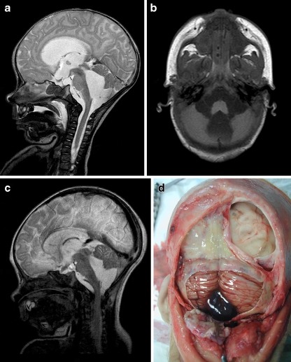
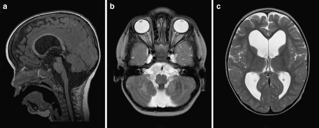
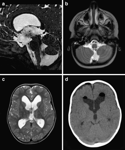
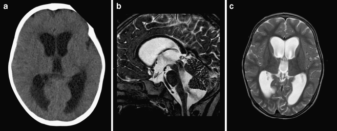
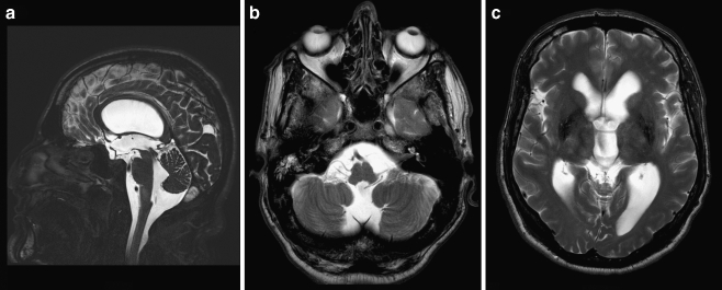
Introduction: Although Blake's pouch cyst (BPC) is frequently mentioned in the spectrum of posterior fossa cysts and cystlike malformations since its first description in 1996, its natural history, clinical presentation, specific imaging characteristics, optimal treatment, and outcome are relatively unknown. Consequently, BPC may still be underdiagnosed. We therefore report six cases ranging from a fatal hydrocephalus in a young boy, over an increasing head circumference with or without impaired neurological development in two infants, to a decompensating hydrocephalus at an advanced age.

Discussion: We focus on their radiological uniformity, which should help making the correct diagnosis, and widely variable clinical presentation, which includes adult cases as well. Differentiating BPC from other posterior fossa cysts and cystlike malformations and recognizing the accompanying hydrocephalus are essentially noncommunicating, not only have important implications on clinical management but also on genetic counseling, which is unnecessary in case of BPC. In our experience, endoscopic third ventriculostomy is a safe and effective treatment option, avoiding the risks and added morbidity of open surgery, as well as many shunt-related problems.


Figures

Fig. 1
Fig. 1
a Preoperative sagittal T2-weighted MR image demonstrating severe hydrocephalus with bulging third ventricular floor, downward bending mammillary bodies, and an open aqueduct. The cerebellum is not rotated. A thin vertical line between dilated fourth ventricle and cisterna magna indicates a Blake’s pouch cyst. b Preoperative axial T1-weighted MR image demonstrating a bilateral indentation on the caudomedial cerebellar surface caused by the BPC. c Postoperative T2-weighted MR image demonstrating the decreased size of both third and fourth ventricle. d Image obtained during postmortem examination demonstrating massive cerebral necrosis and hemorrhagic Blake’s pouch cyst with typical mass effect on both caudomedial cerebellar hemispheres
Fig. 2
Fig. 2
a Preoperative sagittal T1-weighted MR image demonstrating marked hydrocephalus with bulging third ventricular floor, downward bending mammillary bodies, and an open aqueduct. The cerebellum is not rotated. A thin vertical line between dilated fourth ventricle and cisterna magna indicates a Blake’s pouch cyst. The inferior lobules of the vermis are flattened. b Preoperative axial T2-weighted MR image demonstrating a bilateral indentation on the caudomedial cerebellar surface similar to Fig 1b. c Preoperative axial T2-weighted MR image demonstrating enlarged lateral ventricles without periventricular hyperintensities
Fig. 3
Fig. 3
ac Preoperative sagittal (a) and axial (b, c) T2-weighted MR images demonstrating moderate tetraventricular hydrocephalus. Note the flow void between fourth ventricle and Blake’s pouch cyst. d Axial CT scan 3 days postoperatively (although at different angle) demonstrating decreased size of the frontal horns
Fig. 4
Fig. 4
a Preoperative axial CT scan demonstrating marked hydrocephalus with periventricular hypointensities suggesting increased intraventricular pressure. b, c Postoperative sagittal and axial T2-weighted MR images obtained 48 h after discontinued external ventricular drainage. Ventricular size is identical but periventricular hypointensities are less pronounced as on preoperative images. Note an obvious flow void between fourth ventricle and Blake’s pouch cyst (no third ventriculostomy performed)
Fig. 5
Fig. 5
ac Preoperative sagittal (a) and axial (c) T2-weighted and postcontrast sagittal T1-weighted (b) MR images demonstrating marked hydrocephalus and continuation of the fourth ventricular plexus into the Blake’s pouch cyst (black and white arrowheads). The thin vertical septum behind the cerebellum separates the Blake’s pouch cyst from the subarachnoid space
Fig. 6
Fig. 6
ac Sagittal (a) and axial (b, c) T2-weighted MR images demonstrating moderate hydrocephalus with bulging third ventricular floor, downward bending mammillary bodies, and an open aqueduct. The cerebellum is not rotated. A thin vertical line between dilated fourth ventricle and cisterna magna indicates a Blake’s pouch cyst

Comment in

References

    1. Arai H, Sato K. Posterior fossa cysts: clinical, neuroradiological and surgical features. Child’s Nerv Syst. 1990;7:156–164. doi: 10.1007/BF00776713. - DOI - PubMed
    1. Barkovich AJ, Kjos BO, Norman D, Edwards MS. Revised classification of posterior fossa cysts and cystlike malformations based on the results of multiplanar MR imaging. AJNR. 1989;10:977–988. - PubMed
    1. Barkovich AJ. Pediatric neuroimaging. 2. New York: Raven; 1994. pp. 177–275.
    1. Calabrò F, Arcuri T, Jinkins JR. Blake’s pouch cyst: an entity within the Dandy–Walker continuum. Neuroradiology. 2000;42:290–295. doi: 10.1007/s002340050888. - DOI - PubMed
    1. Conti C, Lunardi P, Bozzao A, Liccardo G, Fraioli B. Syringomyelia associated with hydrocephalus and Blake’s pouch cyst: case report. Spine. 2003;28(14):E279–E283. doi: 10.1097/00007632-200307150-00031. - DOI - PubMed

Publication types

MeSH terms