

Review
doi: 10.1007/s00234-009-0644-2.
Epub 2010 Mar 3.
Current concepts of polymicrogyria
Affiliations
- PMID: 20198472
- PMCID: PMC2872023
- DOI: 10.1007/s00234-009-0644-2
Item in Clipboard
Review
Current concepts of polymicrogyria
Neuroradiology.
2010 Jun.
Abstract
Polymicrogyria is one of the most common malformations of cortical development. It has been known for many years and its clinical and MRI manifestations are well described. Recent advances in imaging, however, have revealed that polymicrogyria has many different appearances on MR imaging, suggesting that is may be a more heterogeneous malformation than previously suspected. The clinical and imaging heterogeneity of polymicrogyria is explored in this review.
Figures
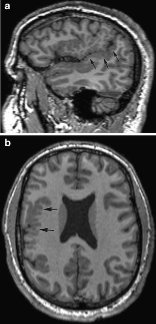
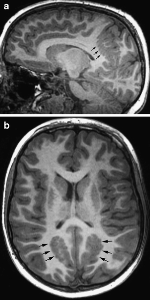
Parasagittal (a) and axial (b) T1-weighted images show the delicate appearance of PMG (black arrows) in the right sylvian and suprasylvian cortex. Note the continuity of the posterior sylvian fissure on the parasagittal image (a); this is diagnostic of perisylvian PMG

Thick and irregularly bumpy, “coarse” appearance of PMG. Parasagittal (a) and axial (b) images show coarse PMG in the parieto-occipital (small black arrows) and perisylvian (large black arrows) regions
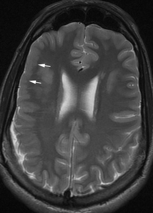
Axial FSE T2 weighted image shows right frontal PMG (white arrows) with fusion of the molecular layer of cortex resulting in paradoxically smooth cortical surface
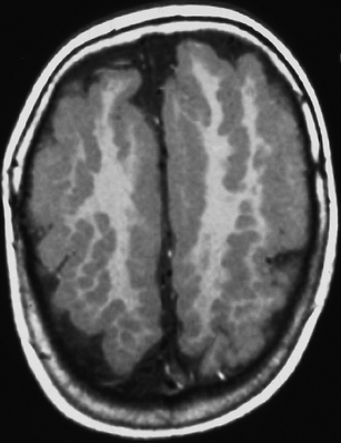
Axial T1-weighted image shows diffuse coarse PMG with an appearance of “palisades” of cortex
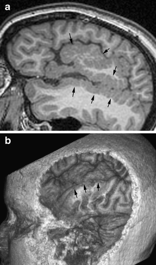
Surface rendering of PMG from volumetric acquisition of data. a Image shows a parasagittal image of extensive perisylvian coarse PMG (arrows). The surface rendering (b) shows the absence of normal sulci and the bumpy cortical surface in the affected area (arrows)
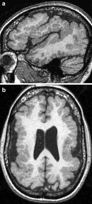
Grade 1 perisylvian PMG. Parasagittal (a) and axial (b) T1-weighted images show coarse polymicrogyria extending from the frontal pole to the occipital pole
Grade 2 perisylvian PMG. Parasagittal (a) and axial (b) images show coarse perisylvian polymicrogyria sparing the anterior frontal lobes and the occipital lobes
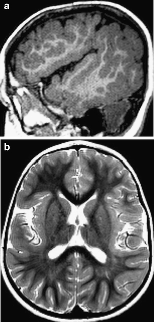
Grade 3 perisylvian PMG. Parasagittal T1-weighted images (a) and axial T2-weighted images (b) show polymicrogyria limited to the insulae and operculae
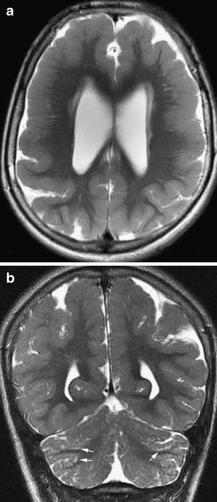
Bilateral frontoparietal polymicrogyria. Axial (a) and coronal (b) T2-weighted images show a different appearance to the cortex, that of multiple radially oriented neuronal components separated by fibro-glial stroma. Note the prominent cerebellar fissures and the subcortical cerebellar cysts (arrows)
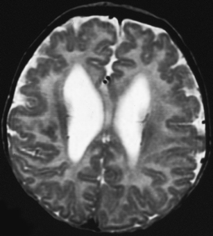
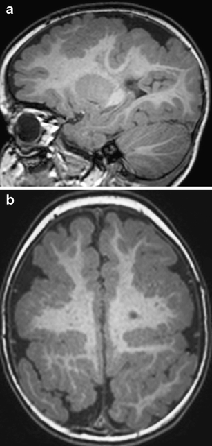
Bilateral frontal polymicrogyria. Axial T2-weighted image shows delicate PMG in both frontal lobes. The lateral ventricles are somewhat dilated
Bilateral parasagittal parieto-occipital polymicrogyria. Parasagittal (a) and axial (b) T1-weighted images show coarse parasagittal infoldings (arrows) of polymicrogyria
Similar articles
-
Cortical thickness reduction of normal appearing cortex in patients with polymicrogyria.J Neuroimaging. 2010 Jan;20(1):46-52. doi: 10.1111/j.1552-6569.2009.00372.x. J Neuroimaging. 2010. PMID: 19453835
-
Compound heterozygosity in GPR56 with bilateral frontoparietal polymicrogyria.Brain Dev. 2014 Jun;36(6):528-31. doi: 10.1016/j.braindev.2013.07.015. Epub 2013 Aug 24. Brain Dev. 2014. PMID: 23981349
-
GPR56-Related Polymicrogyria: Clinicoradiologic Profile of 4 Patients.J Child Neurol. 2015 Nov;30(13):1819-23. doi: 10.1177/0883073815583335. Epub 2015 Apr 28. J Child Neurol. 2015. PMID: 25922261
-
Malformations of cortical development: The role of 7-Tesla magnetic resonance imaging in diagnosis.Rev Neurol (Paris). 2019 Mar;175(3):157-162. doi: 10.1016/j.neurol.2019.01.393. Epub 2019 Mar 1. Rev Neurol (Paris). 2019. PMID: 30827579 Review.
-
Polymicrogyria: a common and heterogeneous malformation of cortical development.Am J Med Genet C Semin Med Genet. 2014 Jun;166C(2):227-39. doi: 10.1002/ajmg.c.31399. Epub 2014 May 28. Am J Med Genet C Semin Med Genet. 2014. PMID: 24888723 Review.
Cited by
-
What disorders of cortical development tell us about the cortex: one plus one does not always make two.Curr Opin Genet Dev. 2011 Jun;21(3):333-9. doi: 10.1016/j.gde.2011.01.006. Epub 2011 Feb 1. Curr Opin Genet Dev. 2011. PMID: 21288712 Free PMC article. Review.
-
Evolutionarily dynamic alternative splicing of GPR56 regulates regional cerebral cortical patterning.Science. 2014 Feb 14;343(6172):764-8. doi: 10.1126/science.1244392. Science. 2014. PMID: 24531968 Free PMC article.
-
Malformations of cortical development: 3T magnetic resonance imaging features.World J Radiol. 2015 Oct 28;7(10):329-35. doi: 10.4329/wjr.v7.i10.329. World J Radiol. 2015. PMID: 26516429 Free PMC article. Review.
-
MR insights into fetal brain development: what is normal and what is not.Pediatr Radiol. 2024 Apr;54(4):635-645. doi: 10.1007/s00247-024-05890-z. Epub 2024 Feb 28. Pediatr Radiol. 2024. PMID: 38416183 Review.
-
Spontaneous Resolution of Congenital Dural Venous Sinus Ectasia Associated With Polymicrogyria-Case Report.Front Pediatr. 2022 Feb 28;10:822551. doi: 10.3389/fped.2022.822551. eCollection 2022. Front Pediatr. 2022. PMID: 35295696 Free PMC article.
References
-
- Norman MG, McGillivray BC, Kalousek DK, Hill A, Poskitt KJ. Congenital malformations of the brain: pathologic, embryologic, clinical, radiologic and genetic aspects. Oxford: Oxford University Press; 1995.
-
- Evrard P, de Saint-Georges P, Kadhim HJ, Gadisseux J-F. Pathology of prenatal encephalopathies. In: French J, editor. Child neurology and developmental disabilities. Baltimore: Paul H. Brookes; 1989. pp. 153–176.
