. 2010 Jun;119(6):755-70.
doi: 10.1007/s00401-010-0655-4. Epub 2010 Mar 3.

The neuropathology of autism: defects of neurogenesis and neuronal migration, and dysplastic changes

Affiliations

The neuropathology of autism: defects of neurogenesis and neuronal migration, and dysplastic changes

Jerzy Wegiel et al. Acta Neuropathol. 2010 Jun.

Abstract

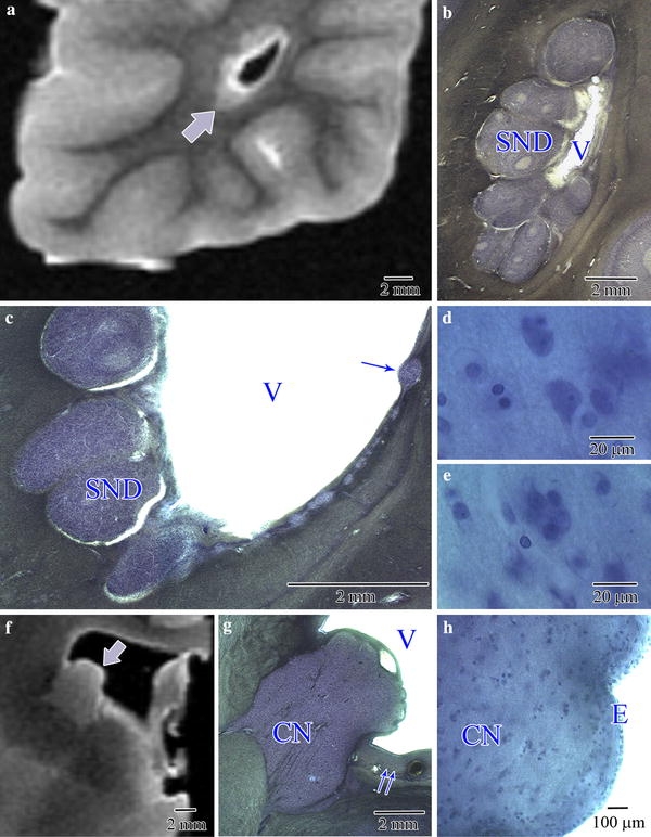
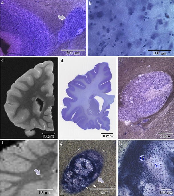
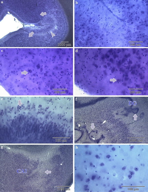
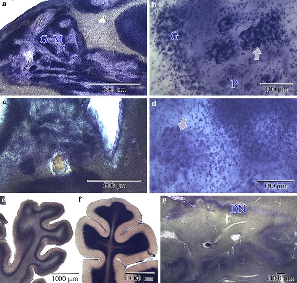
Autism is characterized by a broad spectrum of clinical manifestations including qualitative impairments in social interactions and communication, and repetitive and stereotyped patterns of behavior. Abnormal acceleration of brain growth in early childhood, signs of slower growth of neurons, and minicolumn developmental abnormalities suggest multiregional alterations. The aim of this study was to detect the patterns of focal qualitative developmental defects and to identify brain regions that are prone to developmental alterations in autism. Formalin-fixed brain hemispheres of 13 autistic (4-60 years of age) and 14 age-matched control subjects were embedded in celloidin and cut into 200-mum-thick coronal sections, which were stained with cresyl violet and used for neuropathological evaluation. Thickening of the subependymal cell layer in two brains and subependymal nodular dysplasia in one brain is indicative of active neurogenesis in two autistic children. Subcortical, periventricular, hippocampal and cerebellar heterotopias detected in the brains of four autistic subjects (31%) reflect abnormal neuronal migration. Multifocal cerebral dysplasia resulted in local distortion of the cytoarchitecture of the neocortex in four brains (31%), of the entorhinal cortex in two brains (15%), of the cornu Ammonis in four brains and of the dentate gyrus in two brains. Cerebellar flocculonodular dysplasia detected in six subjects (46%), focal dysplasia in the vermis in one case, and hypoplasia in one subject indicate local failure of cerebellar development in 62% of autistic subjects. Detection of flocculonodular dysplasia in only one control subject and of a broad spectrum of focal qualitative neuropathological developmental changes in 12 of 13 examined brains of autistic subjects (92%) reflects multiregional dysregulation of neurogenesis, neuronal migration and maturation in autism, which may contribute to the heterogeneity of the clinical phenotype.


Figures

Fig. 1
Fig. 1
Nodules in the wall of the lateral ventricle detected in postmortem MRI (a) in the brain of a 7-year-old male diagnosed with autism (B-6403) revealed features of subependymal nodular dysplasia (SND; b) in examination of CV-stained sections. c Numerous large and small nodules (arrow) dispersed within subependymal cell layer. They contained a few pyramidal-like neurons (d) and numerous poorly differentiated cells (e). Tuber-like expansion of the caudate nucleus (arrow) into the ventricle lumen is shown in MRI (f) and in CV-stained section (g). g A thick subependymal cell layer above and below (two arrows) the caudate nucleus (CN), and the absence of matrix in the tuber-like area. Under ependymal (E) cap of the caudate nucleus (CN) tuber-like expansion, small poorly differentiated neurons are present (h)
Fig. 2
Fig. 2
Large subcortical heterotopia within anterior cingulate gyrus in a 5-year-old autistic child (UMB-1627) (a) contained dysplastic neurons without spatial orientation (b). Periventricular heterotopia near the frontal horn of the lateral ventricle (c MRI, d, e CV-stained section) shows a structure resembling molecular, granule and pyramidal layers in a 7-year-old autistic subject (B-6403). MRI (f), low (g) and large (h) magnification of heterotopia (arrow) with dysplastic granule (G) and molecular layer (M) detected within cerebellar white matter in an 11-year-old autistic subject (B-5342)
Fig. 3
Fig. 3
Dysplastic changes within neocortex (a, b), entorhinal cortex (c, d), dentate gyrus (e, f) and the cornu Ammonis (g, h). Focal dysplasia in frontal cortex with loss of vertical and horizontal cytoarchitecture (two arrows) and abnormal (arrowhead) laminar organization (a). Dysplastic neurons within affected area (B-6212) (b). Microdysgenesis within the entorhinal cortex with deficit of stellate neurons in the islands (c) and normal morphology of islands in adjacent cortex (d) in 60-year-old autistic subject (B-7090). Microdysgenesis of the dentate gyrus with dispersion of granule cells within the molecular layer (e, arrow) and distortion of the granule cell layer shape (f, arrows) in 13-year-old autistic male (B-5535). CA1 sector microdysgenesis with local deficit of pyramidal neurons (g, arrow) without markers of gliosis but with signs of poor differentiation of dysplastic abnormally arranged neurons (h) in 13-year-old autistic subject (B-5535)
Fig. 4
Fig. 4
Flocculonodular dysplasia in cerebellum of 56-year-old autistic subject (B-6276) (a) with thin irregular granule (G) and molecular (M) layer. b Dysplastic granule layer (G), ectopic granule cells (arrow) in the molecular layer, and loosely dispersed Purkinje cells (P) (B-6276). Cortical dysplasia within vermis of 13-year-old autistic male (c) with dysplastic granule neurons mixed with heterotopic (arrow) large cells (d) (B-5535). e Severe hypoplasia of cerebellar lobe 3 and unmodified lobe 6 (f), respectively, within the cerebellum of a 60-year-old autistic male (B-7090). In the affected region, the thickness of the hypoplastic molecular and granule cell layer was reduced by about 50%. Almost half of the dentate nucleus (DN) was less convoluted than the unaffected part (g)

Similar articles

Cited by

References

    1. American Psychiatric Association . Diagnostic and statistical manual of mental disorders DSM-IV-TR. Washington, DC: American Psychiatric Association; 2000.
    1. Andrade DM. Genetic basis in epilepsies caused by malformations of cortical development and in those with structurally normal brain. Hum Genet. 2009;126:173–193. - PubMed
    1. Avila J, Dominguez J, Diaz-Nido J. Regulation of microtubule dynamics by microtubule-associated protein expression and phosphorylation during neuronal development. Int J Dev Biol. 1994;38:13–25. - PubMed
    1. Bailey AP, Luthert A, Dean B, et al. A clinicopathological study of autism. Brain. 1998;121:889–905. - PubMed
    1. Bailey AR, Giunta BN, Obregon D, et al. Peripheral biomarkers in autism: secreted amyloid precursor protein-alpha as a probable key player in early diagnosis. Int J Clin Exp Med. 2008;1:338–344. - PMC - PubMed

Publication types