

NAD+ auxotrophy is bacteriocidal for the tubercle bacilli
- PMID: 20199601
- PMCID: PMC2945688
- DOI: 10.1111/j.1365-2958.2010.07099.x
NAD+ auxotrophy is bacteriocidal for the tubercle bacilli
Abstract
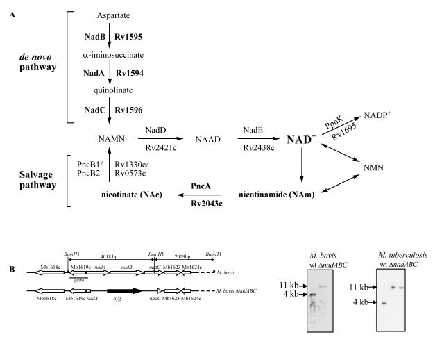
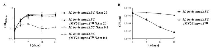
The human tubercle bacillus Mycobacterium tuberculosis can synthesize NAD(+) using the de novo biosynthesis pathway or the salvage pathway. The salvage pathway of the bovine tubercle bacillus Mycobacterium bovis was reported defective due to a mutation in the nicotinamidase PncA. This defect prevents nicotinic acid secretion, which is the basis for the niacin test that clinically distinguishes M. bovis from M. tuberculosis. Surprisingly, we found that the NAD(+)de novo biosynthesis pathway (nadABC) can be deleted from M. bovis, demonstrating a functioning salvage pathway. M. bovisDeltanadABC fails to grow in mice, whereas M. tuberculosisDeltanadABC grows normally in mice, suggesting that M. tuberculosis can acquire nicotinamide from its host. The introduction of M. tuberculosis pncA into M. bovisDeltanadABC is sufficient to fully restore growth in a mouse, proving that the functional salvage pathway enables nicotinamide acquisition by the tubercle bacilli. This study demonstrates that NAD(+) starvation is a cidal event in the tubercle bacilli and confirms that enzymes common to the de novo and salvage pathways may be good drug targets.
Figures


Similar articles
-
Biosynthesis and recycling of nicotinamide cofactors in mycobacterium tuberculosis. An essential role for NAD in nonreplicating bacilli.J Biol Chem. 2008 Jul 11;283(28):19329-41. doi: 10.1074/jbc.M800694200. Epub 2008 May 19. J Biol Chem. 2008. PMID: 18490451 Free PMC article.
-
Evolutionary history of tuberculosis shaped by conserved mutations in the PhoPR virulence regulator.Proc Natl Acad Sci U S A. 2014 Aug 5;111(31):11491-6. doi: 10.1073/pnas.1406693111. Epub 2014 Jul 21. Proc Natl Acad Sci U S A. 2014. PMID: 25049399 Free PMC article.
-
Metabolism of nicotinamide adenine dinucleotide in human and bovine strainsof Mycobacterium tuberculosis.J Bacteriol. 1972 May;110(2):600-3. doi: 10.1128/jb.110.2.600-603.1972. J Bacteriol. 1972. PMID: 4336690 Free PMC article.
-
Mycobacterium bovis lipids: virulence and vaccines.Vet Microbiol. 2011 Jul 5;151(1-2):91-8. doi: 10.1016/j.vetmic.2011.02.030. Epub 2011 Feb 24. Vet Microbiol. 2011. PMID: 21420803 Review.
-
Genomics of Mycobacterium bovis.Tuberculosis (Edinb). 2001;81(1-2):157-63. doi: 10.1054/tube.2000.0269. Tuberculosis (Edinb). 2001. PMID: 11463237 Review.
Cited by
-
Comprehensive insights into transcriptional adaptation of intracellular mycobacteria by microbe-enriched dual RNA sequencing.BMC Genomics. 2015 Feb 5;16(1):34. doi: 10.1186/s12864-014-1197-2. BMC Genomics. 2015. PMID: 25649146 Free PMC article.
-
Perspectives of Hospital Staff on Barriers to Smoking Cessation Interventions among Drug-Resistant Tuberculosis Patients in a South African Management Hospital.Int J Environ Res Public Health. 2024 Aug 28;21(9):1137. doi: 10.3390/ijerph21091137. Int J Environ Res Public Health. 2024. PMID: 39338021 Free PMC article.
-
The Bewildering Antitubercular Action of Pyrazinamide.Microbiol Mol Biol Rev. 2020 Mar 4;84(2):e00070-19. doi: 10.1128/MMBR.00070-19. Print 2020 May 20. Microbiol Mol Biol Rev. 2020. PMID: 32132245 Free PMC article. Review.
-
Pantothenate and pantetheine antagonize the antitubercular activity of pyrazinamide.Antimicrob Agents Chemother. 2014 Dec;58(12):7258-63. doi: 10.1128/AAC.04028-14. Epub 2014 Sep 22. Antimicrob Agents Chemother. 2014. PMID: 25246400 Free PMC article.
-
Critical role for NLRP3 in necrotic death triggered by Mycobacterium tuberculosis.Cell Microbiol. 2011 Sep;13(9):1371-84. doi: 10.1111/j.1462-5822.2011.01625.x. Epub 2011 Jul 11. Cell Microbiol. 2011. PMID: 21740493 Free PMC article.
References
-
- Bardarov S, Bardarov S, Jr, Pavelka MS, Jr, Sambandamurthy V, Larsen M, Tufariello J, Chan J, Hatfull G, Jacobs WR., Jr Specialized transduction: an efficient method for generating marked and unmarked targeted gene disruptions in Mycobacterium tuberculosis, M. bovis BCG and M. smegmatis. Microbiology. 2002;148:3007–3017. Jr. Jr. Jr. - PubMed
-
- Begley TP, Kinsland C, Mehl RA, Osterman A, Dorrestein P. The biosynthesis of nicotinamide adenine dinucleotides in bacteria. Vitam. Horm. 2001;61:103–119. - PubMed
-
- Bekierkunst A, Artman M. Tissue metabolism in infection. DPNase activity, DPN levels, and DPN-linked dehydrogenases in tissues from normal and tuberculous mice. Am. Rev. Respir. Dis. 1962;86:832–838. - PubMed
-
- Betts JC, Lukey PT, Robb LC, McAdam RA, Duncan K. Evaluation of a nutrient starvation model of Mycobacterium tuberculosis persistence by gene and protein expression profiling. Mol Microbiol. 2002;43:717–731. - PubMed
-
- Boshoff HI, Myers TG, Copp BR, McNeil MR, Wilson MA, Barry CE., 3rd The transcriptional responses of Mycobacterium tuberculosis to inhibitors of metabolism: novel insights into drug mechanisms of action. J Biol Chem. 2004;279:40174–40184. - PubMed
Publication types
MeSH terms
Substances
Grants and funding
LinkOut - more resources
Full Text Sources
Other Literature Sources
Molecular Biology Databases
