. 2010 Apr;76(1):236-59.
doi: 10.1111/j.1365-2958.2010.07096.x. Epub 2010 Feb 28.

A dominant-negative needle mutant blocks type III secretion of early but not late substrates in Yersinia

Affiliations

A dominant-negative needle mutant blocks type III secretion of early but not late substrates in Yersinia

Alison J Davis et al. Mol Microbiol. 2010 Apr.

Abstract

Yersinia pseudotuberculosis uses a type III secretion system (T3SS) to deliver effectors into host cells. A key component of the T3SS is the needle, which is a hollow tube on the bacterial surface through which effectors are secreted, composed of the YscF protein. To study needle assembly, we performed a screen for dominant-negative yscF alleles that prevented effector secretion in the presence of wild-type (WT) YscF. One allele, yscF-L54V, prevents WT YscF secretion and needle assembly, although purified YscF-L54V polymerizes in vitro. YscF-L54V binds to its chaperones YscE and YscG, and the YscF-L54V-EG complex targets to the T3SS ATPase, YscN. We propose that YscF-L54V stalls at a binding site in the needle assembly pathway following its release from the chaperones, which blocks the secretion of WT YscF and other early substrates required for building a needle. Interestingly, YscF-L54V does not affect the activity of pre-assembled actively secreting machines, indicating that a factor and/or binding site required for YscF secretion is absent from T3SS machines already engaged in effector secretion. Thus, substrate switching may involve the removal of an early substrate-specific binding site as a mechanism to exclude early substrates from Yop-secreting machines.


Figures

Figure 1
Figure 1
The yscF L54V mutant prevents secretion of WT YscF, needle formation and secretion of Yops. WT or ΔyscF strains carrying plasmids pTRC99-yscF, pTRC99A or pTRC99-yscF-L54V were grown in low calcium media and shifted to 37°C. Expression of yscF or yscF-L54V from the plasmids was induced with IPTG at the 37°C temperature shift. Cell-associated and secreted proteins harvested from equal numbers of bacteria were separated by SDS-PAGE and detected by coomassie staining or by Western blotting. A. No Yops or YscF are secreted in the presence of yscF-L54V. Secreted proteins visualized by coomassie staining (top panel). Secreted (middle panel) and cell-associated (lower panel) YscF levels detected by Western blot with α-YscF antiserum. B. YscF-L54V protein does not make needles and prevents WT YscF needle formation. Equal numbers of bacteria were collected and exposed to water or the chemical cross-linker BS3 (−/+ BS3). Bacteria were solubilized in SDS-sample buffer and proteins were separated by SDS-PAGE. YscF protein was detected by Western blotting with antibodies to YscF. YscF monomers are ~ 7 kDa and YscF polymers run as a ladder of higher molecular weight bands (polymers). Non-specific antibody-reactive bands are apparent in the no-cross-linker control samples (odd numbered lanes). C. Expression of yscF-L54V decreases levels of T3SS-associated proteins. Cellular levels of YopE, YopD and YscN were visualized by Western blotting. Molecular mass markers are shown on the left in kDa in A and B.
Figure 2
Figure 2
yscF-L54V expression prevents needle assembly during growth in high calcium media. WT or ΔyscF strains carrying plasmids pTRC99-yscF, pTRC99A or pTRC99-yscF-L54V were grown in media supplemented with 3 mM calcium and shifted to 37°C. Expression of yscF from plasmids was induced with IPTG at the 37°C temperature shift. A. Expression of yscF L54V in high calcium does not affect cellular levels of T3SS-associated proteins. YscF, YopE, YopD and YscN proteins were visualized by Western blotting. B. No external YscF polymers are present in yscF L54V-containing strains. Equal numbers of bacteria were cross-linked with BS3 and processed as in Figure 1B. YscF protein was detected by Western blotting. C. No external YscF protein is present in YscF-L54V-containing strains. Bacteria were fixed without permeabilization, adhered to glass coverslips, and stained with antibodies to YscF followed by Alexa592 secondary antibodies (red) as described in the Experimental Procedures. DAPI (blue) was used to detect nucleoids. Samples were visualized by fluorescence microscopy.
Figure 3
Figure 3
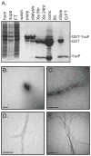
YscF-L54V protein polymerizes in vitro. A. Recombinant YscF protein purification profile. Commassie stained gel of samples taken during the purification of YscF (see Experimental Procedures for details). YscF (shown) and L54V proteins were over-expressed in E. coli as GST fusions, purified from the soluble fraction (supe) using glutathione-agarose, and eluted with free glutathione (elute). Following dialysis to remove the glutathione (dialysis), YscF was released from GST by Factor Xa cleavage (Xa). The GST and YscF mixture was concentrated (conc.) and diluted into 10 mM Tris pH 8.0 (dilute). GST was removed with Q sepharose (Q FT), leaving free YscF. Purification of the YscF-L54V protein proceeded identically. B–E. WT YscF and YscF-L54V proteins polymerize in vitro. Purified YscF (B, C) or YscF-L54V (D, E) proteins were mounted onto carbon-coated EM grids and negatively stained with uranyl acetate immediately after purification (B) or after incubation at room temperature for 16 hrs (C–E). Samples were visualized by transmission electron microscopy. Bars are 200 nm (B–D) and 500 nm (E).
Figure 4
Figure 4
Residue L54 resides in the YscF-YscG hydrophobic pocket but does not disrupt complex formation. A. Location of YscF residue L54 within the YscF-EG complex. The crystal structure model of the YscF-EG complex is shown; YscG is in grey with the α-1 chain labeled, YscE is in violet and YscF is in green. The leucine 54 side chain of YscF is highlighted in red. Figure was prepared using PyMol (DeLano, 2008). B. YscF-L54V forms a complex with YscE and YscG in Yersinia. Virulence plasmid- cured Yersinia strains (pYV-) containing pFlag-YscG-YscE-His and either pBAD-yscF or pBAD-yscF-L54V were induced to co-express Flag-YscG, YscE-His and either YscF or YscF-L54V by addition of IPTG and arabinose. Bacteria were harvested, lysed, and the soluble fraction (start) was precipitated with Flag antibody-conjugated agarose beads. The flow-through (FT) fraction was collected, and the beads were washed thoroughly (wash). Co-precipitated protein complexes were eluted by boiling in SDS sample buffer (elute). Proteins in the different fractions were separated by SDS-PAGE, and detected by Western blot with antibodies to Flag (YscG), His (YscE) and YscF.
Figure 5
Figure 5
Over-expression of WT YscF-EG proteins suppresses YscF-L54V DN phenotypes. WT or yscF-L54V strains containing pBAD33 (lanes 1 and 5), pBAD-yscF (lanes 2 and 6), pFlag-YscG-YscE-His and pBAD33 (lanes 3 and 7) or pFlag-YscG-YscE-His and pBAD-yscF (lanes 4 and 8) were grown in low-calcium media and expression from plasmids was induced at the 37°C temperature shift by addition of arabinose, IPTG, or both. Bacteria and culture supernatants were collected and proteins separated by SDS-PAGE. A. Secreted Yops detected by coomassie staining. B. Cell-associated (cells) and secreted YscF protein from above strains detected by Western blot with α-YscF antibodies. C. Above strains were chemically cross-linked with BS3 (lanes 1–8). Cross-linked samples and non-cross-linked WT bacteria (BS3) were solubilized in SDS-sample buffer and separated by SDS-PAGE. YscF monomer and polymers were visualized by Western blot with antibodies to YscF. Molecular mass markers for A and C are shown on the left in kDa.
Figure 6
Figure 6
Purification and biochemical analysis of YscF-EG and YscF-L54V-EG complexes. A. Gel filtration elution traces of WT YscF-EG and L54V-EG complexes. Flag-affinity purification eluates were passed over a Sephacryl S-100 HR gel filtration column. Five peaks were observed (P1–P5) for YscF-EG (blue) and YscF-L54V-EG (red) samples. The Y-axis shows the absorbance at 280 nm. The X-axis shows the elution volume in ml. The column was calibrated with albumin (67 kDa), ovalbumin (43 kDa), chymotrypsinogen A (25 kDa), and Ribonuclease A (13.7 kDa). B. The eluate and peak fractions (P1–P4) from A were concentrated, proteins were separated by SDS-PAGE, and protein composition in peak samples was analyzed by coomassie staining. C. Peak 2 and peak 4 samples from both YscF-EG (WT) and YscF-L54V-EG (L54V) were analyzed by native PAGE and Western blotting. Migration as a complex (closed arrowhead) or a monomer (open arrowhead) is indicated. D. Protease protection assay. Three μg of YscF-EG or YscF-L54V-EG peak 2 complexes (lanes 1 and 7) was incubated with 1 μg, 0.2μg, 0.04 μg or 0.008 μg of thermolysin (lanes 2–5, and 8–11) or with 1 μg of thermolysin and 1% TritonX-100 (lanes 6 and 12) for 1 hr. at 37°C. Proteins were separated by SDS-PAGE and stained with coomassie. Full-length Flag-YscG (G), YscE-His (E) and YscF (F) proteins are labeled. E. Thermal denaturation curves. Purified YscF-EG (blue lines), YscF-L54V-EG complexes (red lines) and the P4 fraction from the WT YscF-EG purification in Fig 6A (black line) were exposed to a temperature gradient of 1°C per min in the presence of the dye Sypro Orange. Fluorescence intensity was monitored as the temperature increased. The fluorescence minimum and maximum for each sample was set to 0 and 1, respectively, and the normalized data are shown as relative fluorescence. Needle-chaperone complex samples were tested in triplicate and all curves are shown.
Figure 7
Figure 7
A. YscN physically interacts with both the YscF-EG and YscF-L54V-EG complexes in Yersinia. WT, yscF L54V or ΔyscF strains expressing Flag-YscG, YscE-His and His-YscN, as well as control WT strains expressing either Flag-YscG and YscE-His or His-YscN were grown in low-calcium media at 37°C. Bacteria were lysed and insoluble protein was removed by centrifugation (P). Clarified supernatants (S) were passed over a Flag affinity column, washed (wash), and proteins eluted by competition with the Flag peptide (elute). Samples were separated by SDS-PAGE and analyzed by Western blotting with antibodies to Flag (YscG), His (YscE and YscN), and YscF. Arrowheads indicate location of YscN protein in the eluates. B. Excess YscN protein does not suppress the yscF L54V secretion defect. WT Yersinia containing the plasmids pTRC99 and pBAD33 (lane 1), pTRC99 and pBAD-yscF L54V (lane 2), pTRC99-his yscN and pBAD33 (lane 3), or pTRC99-his yscN and pBAD-yscF L54V (lane 4) were grown in low-calcium media, and expression from plasmids was induced at the 37°C shift. After 2 hrs at 37°C, cell pellets and culture supernatants were collected and proteins were separated by SDS-PAGE. Secreted Yops in culture supernatants were visualized by Coomassie staining (top panel). Proteins in cell pellets were visualized by western blotting with antibodies to YscN, the His tag, and YscF (middle three panels). Secreted YscF was detected in culture supernatants with YscF antibodies (bottom panel). C. YscF-EG and YscF-L54V-EG complexes stimulate equal levels of YscN ATPase activity. Phosphate release was measured to detect ATPase activity of purified His-YscN protein in the presence of ATP, and when mixed with purified YscF-EG or YscF-L54V-EG complexes and ATP. Shown is a representative experiment, performed in duplicate. Error bars indicate the standard error of the mean.
Figure 8
Figure 8
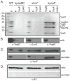
yscF-L54V blocks Yop and YscF secretion in regulation-defective strain backgrounds. Strains deleted for yopB and yopDyopBD), lcrVlcrV), or yopNyopN) carrying plasmids pTRC99A, pTRC99-yscF or pTRC99-yscF-L54V were grown in low calcium media and shifted to 37°C. Cell-associated and secreted proteins were processed as in Figure 1A. A. Secreted proteins visualized by coomassie staining. Molecular mass standards are shown o the left in kDa. Yops are labeled on the right. B. Cellular YopD (left panel), LcrV (middle panel) and YopN (right panel) proteins in the corresponding deletion strains visualized by Western blotting. C. Cellular (top panel) and secreted (bottom panel) YscF protein visualized by Western blotting. D. Cytosolic control protein S2 visualized by Western blotting in the cellular (top panel) and secreted (bottom panel) protein fractions.
Figure 9
Figure 9
YscF-L54V does not block Yop secretion from preformed WT secreting needles. The ΔyscF+pBAD-yscF, WT+pBAD and WT+pBAD-yscF-L54V strains were grown at 26°C in high or low calcium media in the presence of dextrose to repress expression from the pBAD promoter. Cultures were shifted to 37°C for 30 minutes to allow for production of either non-secreting needles or secreting needles in the WT backgrounds. Samples were then split in half, cells were washed to remove the dextrose and moved into low calcium media pre-warmed to 37°C. Expression from the pBAD plasmids either remained repressed with dextrose, or was induced by addition of arabinose and incubated at 37°C for 30 minutes (panels 3–6). Media was then removed from all cultures and was replaced with pre-warmed fresh media of the same composition and growth continued at 37°C for an additional 60 minutes (panels 7–14). Samples were taken at various steps and processed for immunofluorescence and secreted Yops. A. Immunofluorescence micrographs of surface-localized YscF on the WT+pBAD-yscF-L54V strain (panels 1–10) and the WT+pBAD strain (panels 11–14). Experimental schematic is shown on the left. At the indicated steps, samples were removed and bacteria were fixed and stained with DAPI and YscF antibodies as in Figure 2C. B. Secreted Yops were collected from culture supernatants of all three strains that were induced with arabinose or not (+/− pBAD induction) after the final incubation step in A. Proteins were separated by SDS-PAGE and stained with coomassie. Numbers under gel lanes refer to the corresponding samples shown in A, panels 7–14.

Similar articles

Cited by

References

    1. Abe A, de Grado M, Pfuetzner RA, Sanchez-Sanmartin C, Devinney R, Puente JL, Strynadka NC, Finlay BB. Enteropathogenic Escherichia coli translocated intimin receptor, Tir, requires a specific chaperone for stable secretion. Mol Microbiol. 1999;33:1162–1175. - PubMed
    1. Agrain C, Sorg I, Paroz C, Cornelis GR. Secretion of YscP from Yersinia enterocolitica is essential to control the length of the injectisome needle but not to change the type III secretion substrate specificity. Mol Microbiol. 2005;57:1415–1427. - PubMed
    1. Akeda Y, Galan JE. Chaperone release and unfolding of substrates in type III secretion. Nature. 2005;437:911–915. - PubMed
    1. Allaoui A, Schulte R, Cornelis GR. Mutational analysis of the Yersinia enterocolitica virC operon: characterization of yscE, F, G, I, J, K required for Yop secretion and yscH encoding YopR. Mol Microbiol. 1995;18:343–355. - PubMed
    1. Amann E, Ochs B, Abel KJ. Tightly regulated tac promoter vectors useful for the expression of unfused and fused proteins in Escherichia coli. Gene. 1988;69:301–315. - PubMed

Publication types

MeSH terms