. 2010 Jun 1;76(5):1127-41.
doi: 10.1111/j.1365-2958.2010.07092.x. Epub 2010 Feb 28.

Mycoplasma pneumoniae Community Acquired Respiratory Distress Syndrome toxin expression reveals growth phase and infection-dependent regulation

Affiliations
Free PMC article

Mycoplasma pneumoniae Community Acquired Respiratory Distress Syndrome toxin expression reveals growth phase and infection-dependent regulation

T R Kannan et al. Mol Microbiol. .
Free PMC article

Abstract

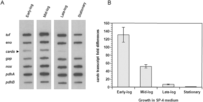
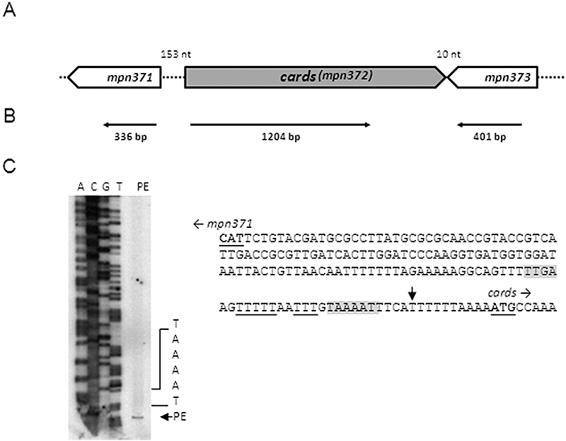
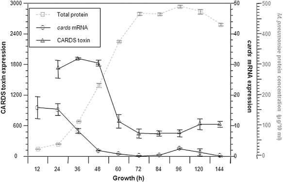
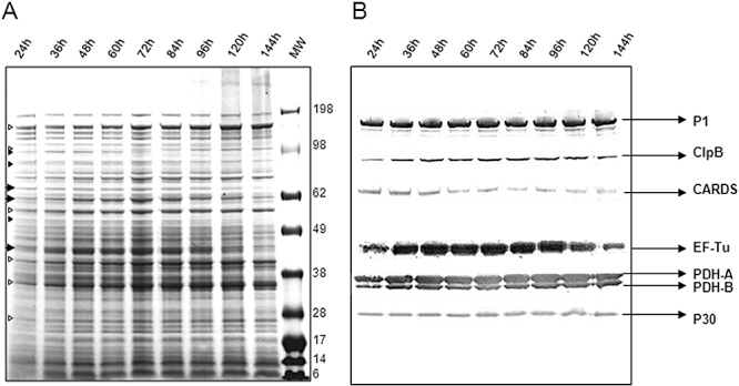
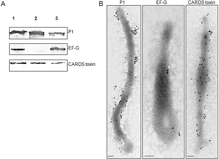
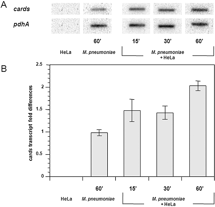
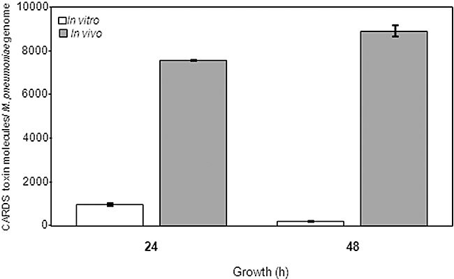
Mycoplasma pneumoniae causes acute and chronic respiratory infections, including tracheobronchitis and community acquired pneumonia, and is linked to asthma and an array of extra-pulmonary disorders. Recently, we identified an ADP-ribosylating and vacuolating toxin of M. pneumoniae, designated Community Acquired Respiratory Distress Syndrome (CARDS) toxin. In this study we analysed CARDS toxin gene (annotated mpn372) transcription and identified its promoter. We also compared CARDS toxin mRNA and protein profiles in M. pneumoniae during distinct in vitro growth phases. CARDS toxin mRNA expression was maximal, but at low levels, during early exponential growth and declined sharply during mid-to-late log growth phases, which was in direct contrast to other mycoplasma genes examined. Between 7% and 10% of CARDS toxin was localized to the mycoplasma membrane at mid-exponential growth, which was reinforced by immunogold electron microscopy. No CARDS toxin was released into the medium. Upon M. pneumoniae infection of mammalian cells, increased expression of CARDS toxin mRNA was observed when compared with SP-4 broth-grown cultures. Further, confocal immunofluorescence microscopy revealed that M. pneumoniae readily expressed CARDS toxin during infection of differentiated normal human bronchial epithelial cells. Analysis of M. pneumoniae-infected mouse lung tissue revealed high expression of CARDS toxin per mycoplasma cell when compared with M. pneumoniae cells grown in SP-4 medium alone. Taken together, these studies indicate that CARDS toxin expression is carefully controlled by environmental cues that influence its transcription and translation. Further, the acceleration of CARDS toxin synthesis and accumulation in vivo is consistent with its role as a bona fide virulence determinant.


Figures

Fig. 2
Fig. 2
Transcription of cards during in vitro growth of M. pneumoniae in SP-4 broth. A. Expression of cards along with other genes was analysed by DNA slot blot. M. pneumoniae gene-specific PCR products (Table S2) were blotted onto Zeta probe membranes. M. pneumoniae S1 cells were grown and harvested at early-log (24 h), mid-log (48 h), late-log (72 h) and stationary phases (120 h). 32P-labelled cDNAs generated by reverse transcription of isolated total RNA were used as hybridization probes. Experiments were repeated three times. B. Expression of cards by relative real-time quantitative RT-PCR (qRT-PCR). M. pneumoniae S1 cells were grown and RNA isolated at specific growth phases. Real-time qRT-PCR was performed using SYBR green chemistry as detailed in Experimental procedures. Experiments were repeated two times. The average fold differences in expression levels of cards mRNA at various time points (when compared with the stationary phase) and standard deviations (SD) are presented.
Fig. 1
Fig. 1
Chromosomal organization and transcriptional and primer extension analyses of cards (mpn372). A. Schematic of cards and its surrounding genes. mpn371 and mpn373 encode hypothetical proteins. The orientation of individual genes is indicated (open arrows), and numbers represent the length of intergenic regions. B. Transcriptional analysis. RT-PCR was performed on total RNA isolated from M. pneumoniae cells. The thin horizontal arrows represent regions that were amplified to confirm transcription of individual genes, and numbers represent sizes of RT-PCR amplified products. C. Primer extension (PE) analysis and characterization of cards gene promoter. The PE product was separated by electrophoresis and analysed alongside DNA sequence (A, T, C and G). The sequence of the intergenic region separating mpn371 and cards is provided. The translational start sites of mpn371 and cards are indicated (bold letters). A vertical arrow designates the transcriptional start point identified for cards. Putative Pribnow box (−10 site), along with M. pneumoniae conserved TTGA sequences, are shaded in gray colour. Poly-T tracts are underlined.
Fig. 4
Fig. 4
Growth phase-dependent variations in cards mRNA and CARDS toxin protein levels in M. pneumoniae broth cultures. M. pneumoniae cells were grown at 37°C for 12, 24, 36, 48, 60, 72, 84, 96 and 120 h and harvested at each time interval for analysis. Experiments were repeated two times. Levels of cards transcript are estimated by DNA slot blot (average value ± SD), CARDS toxin protein levels are quantified by immunoblot, and mycoplasma growth (total protein) is indicated in grey dotted lines (average value ± SD), and expression levels are presented in arbitrary values.
Fig. 3
Fig. 3
Differential synthesis of M. pneumoniae proteins during in vitro growth. M. pneumoniae S1 cells were grown in SP-4 broth for various times and samples collected and processed as described in Experimental procedures. A. Equal amounts of S1 total cell proteins resolved on 4–12% Nu-PAGE gradient gel and stained with Coomassie Blue. Open arrows: increased protein intensity during early-to-late growth stages. Small closed arrows: decreased protein intensity during mid-to-late growth stages. Large closed arrows: increased protein intensity during stationary stage. B. Parallel gel transferred to nitrocellulose membrane for immunoblotting. Membrane strips were cut and treated with respective M. pneumoniae antibodies. Experiments were repeated two times.
Fig. 5
Fig. 5
Cell fractionation of M. pneumoniae and localization of CARDS toxin. A. Total lysate, membrane and cytoplasmic fractions of M. pneumoniae cells were isolated as described in Experimental procedures. Equal amounts of mycoplasma proteins from each fraction were resolved on 4–12% gradient Nu-PAGE gels and transferred to nitrocellulose membranes. Specific proteins were identified by immunoblotting for the presence of CARDS toxin, cytoplasmic EF-G and membrane adhesin P1. Lane 1. total cell lysate; lane 2. membrane fraction; and lane 3. cytoplasmic fraction. B. Immunogold electron microscopy detection of CARDS toxin and control proteins P1 and EF-G on intact M. pneumoniae cells. M. pneumoniae cells were gold particle-labelled with anti-P1, anti-EF-G and anti-CARDS toxin antibodies as described in Experimental procedures. Mycoplasma cells were visualized with a JEOL 1230 transmission electron microscope. Gold labelling of CARDS toxin revealed surface membrane distribution throughout the M. pneumoniae membrane and tip organelle (bar = 0.1 µm). Experiments were repeated two times.
Fig. 7
Fig. 7
Localization of CARDS toxin in M. pneumoniae-infected NHBE cells. Fully differentiated NHBE cells grown in air–liquid interface were infected with M. pneumoniae. Thirty-eight hours post infection, cultures were washed to remove unbound mycoplasmas, submerged in fixative, processed for laser scanning confocal microscopy and screened using mouse polyclonal anti-CARDS toxin sera. Luminal (A1 and B1) and cross-sectional (A2 and B2) views of uninfected (A1 and A2) and infected (B1 and B2) NHBE cells are presented. In the latter case, surface colonization by mycoplasmas and synthesis and distribution of CARDS toxin during infection are readily observed (red). Cross-sectional views of infected cells reveal intracellular localization of CARDS toxin (arrows). Also, DAPI staining indicates the abundance of surface associated M. pneumoniae DNA (small blue dots on the respiratory cell surface; B1 and B2). Merged images of the cross-sectional view indicate colocalization of CARDS toxin with M. pneumoniae at respiratory cell surfaces and also demonstrate that toxin is detectable inside target cells, possibly free of associated M. pneumoniae. Experiments were repeated three times.
Fig. 6
Fig. 6
Differential expression of cards mRNA in M. pneumoniae during co-incubation with HeLa cells. A. Autoradiographic DNA slot blot analysis for cards and pdhA genes. M. pneumoniae-specific primers were used to generate 32P-labelled cDNAs from total RNA isolated from HeLa cells alone, M. pneumoniae cells alone or M. pneumoniae cells co-incubated with HeLa cells for different time intervals. Experiments were repeated three times. B. Relative quantification of cards mRNA by real-time qRT-PCR. M. pneumoniae S1 cells were co-incubated with HeLa cells for 15, 30 and 60 min as described in Experimental procedures, and RNA was isolated. Real-time qRT-PCR was performed using SYBR green chemistry and pdhA as a normalizer. Experiments were repeated two times. The average fold differences in expression levels of cards mRNA at various time points (when compared with the cards mRNA level at time 0) and SD are presented.
Fig. 8
Fig. 8
Differential expression of CARDS toxin in mice. Mice were infected intranasally with M. pneumoniae, and lungs were harvested after 24 and 48 h of infection and analysed for the presence of CARDS toxin molecules per M. pneumoniae cell by antigen capture assay and quantitative real-time PCR (qPCR). In parallel, M. pneumoniae cells were grown in SP-4 broth, harvested and analysed similarly. Numbers of CARDS toxin molecules per mycoplasma genome in SP-4-grown M. pneumoniae cells (in vitro) or in lungs of infected mice (in vivo) are represented by bar diagrams. Experiments were repeated two times with three to five animals of each group and at each time point.

Similar articles

Cited by

References

    1. Balasubramanian S, Kannan TR, Baseman JB. The surface-exposed carboxyl region of Mycoplasma pneumoniae elongation factor Tu interacts with fibronectin. Infect Immun. 2008;76:3116–3123. - PMC - PubMed
    1. Barile MF, Chandler DK, Yoshida H, Grabowski MW, Harasawa R, Ahmed OA. Hamster challenge potency assay for evaluation of Mycoplasma pneumoniae vaccines. Isr J Med Sci. 1981;17:682–686. - PubMed
    1. Baseman JB, Tully JG. Mycoplasmas: sophisticated, reemerging, and burdened by their notoriety. Emerg Infect Dis. 1997;3:21–32. - PMC - PubMed
    1. Baseman JB, Lange M, Criscimagna NL, Giron JA, Thomas CA. Interplay between mycoplasmas and host target cells. Microb Pathog. 1995;19:105–116. - PubMed
    1. Chang LJ, Chen WH, Minion FC, Shiuan D. Mycoplasmas regulate the expression of heat-shock protein genes through CIRCE-HrcA interactions. Biochem Biophys Res Commun. 2008;367:213–218. - PubMed

Publication types

MeSH terms