Review
. 2010 Mar;90(2):84-93.
doi: 10.1016/j.tube.2010.02.003. Epub 2010 Mar 3.

Diversity in Mycobacterium tuberculosis mannosylated cell wall determinants impacts adaptation to the host

Affiliations
Review

Diversity in Mycobacterium tuberculosis mannosylated cell wall determinants impacts adaptation to the host

Jordi B Torrelles et al. Tuberculosis (Edinb). 2010 Mar.

Abstract

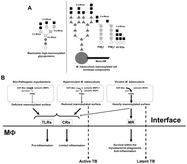
Mycobacterium tuberculosis (the causal agent of TB) has co-evolved with humans for centuries. It infects via the airborne route and is a prototypic highly adapted intracellular pathogen of macrophages. Extensive sequencing of the M. tuberculosis genome along with recent molecular phylogenetic studies is enabling us to gain insight into the biologic diversity that exists among bacterial strains that impact the pathogenesis of latent infection and disease. The majority of the M. tuberculosis cell envelope is comprised of carbohydrates and lipids, and there is increasing evidence that these microbial determinants that are readily exposed to the host immune system play critical roles in disease pathogenesis. Studies from our laboratory and others have raised the possibility that M. tuberculosis is adapting to the human host by cloaking its cell envelope molecules with terminal mannosylated (i.e. Man-alpha-(1-->2)-Man) oligosaccharides that resemble the glycoforms of mammalian mannoproteins. These mannosylated biomolecules engage the mannose receptor (MR) on macrophages during phagocytosis and dictate the intracellular fate of M. tuberculosis by regulating formation of the unique vesicular compartment in which the bacterium survives. The MR is highly expressed on alveolar macrophages (predominant C-type lectin on human cells) and functions as a scavenger receptor to maintain the healthiness of the lung by clearing foreign particles and at the same time regulating dangerous inflammatory responses. Thus M. tuberculosis exploits MR functions to gain entry into the macrophage and survive. Key biochemical pathways and mycobacterial determinants involved in the development and maintenance of the M. tuberculosis phagosome are being identified. The phylogenetic diversity observed in M. tuberculosis strains that impact its cell wall structure together with the genetic diversity observed in human populations, including those elements that affect macrophage function, may help to explain the extraordinary evolutionary adaptation of this pathogen to the human host. Major developments in these areas are the focus of this review.


Conflict of interest statement

Conflict of Interest

The authors have not financial and personal conflict of interest.

Figures

Figure 1
Figure 1. The cell envelope of M. tuberculosis with an emphasis on exposed mannosylated cell envelope components
This scheme depicts the cell envelope “skeleton or core” determinants (mycolyl-arabinogalactan-peptidoglycan complex) and emphasizes the distribution of intercalated major mannosylated cell envelope components that are exposed on the M. tuberculosis surface. AG is covalently linked to PG via the galactan chain and the arabinan chain is in turn linked to the mycolic acids (Myc Ac) which are shown perpendicular to the plasma membrane. The polar groups (i.e. carbohydrate domains) of several mannosylated cell envelope components are exposed on the cell surface and their lipid domains are intercalated with the Myc Ac acid layer. These envelope components include ManLAM, LM, higher- and lower-order PIMs, and lipomannoproteins. Other known virulence factors described for M. tuberculosis that interact with the Myc Ac layer [i.e. TDM, SL; and TGs and PGL, the latter on some M. tuberculosis strains)] are also depicted. Not all Myc Ac are depicted interacting with cell surface components. Not shown are capsule-like components (i.e., arabinomannan, glucan, mannan, and xylan). In order to maintain simplicity, molecular quantities depicted (relative number of molecules) do not accurately reflect experimental data. AG (arabinogalactan); PG (peptidoglycan); Myc Ac (mycolic acids); ManLAM (mannose-capped lipoarabinomannan); LM (lipomannan); PIMs (phosphatidyl-myo-inositol mannosides); TDM (trehalose dimycolate); SL (sulfolipid); TGs (triglycerides); PGL (phenolic glycolipid).
Figure 2
Figure 2. Phagosomes contain ManLAM exposed on their cytosolic face
A. Purified phagosomes containing ManLAM-coated or human serum albumin (HSA)-coated (control) FITC positive beads were incubated with anti-ManLAM (CS-35) or mouse IgG for 20 minutes, washed, stained with secondary anti-mouse IgG -APC and analyzed on a LSRII flow cytometer. A subset of phagosomes was permeabilized (P) using reagents from BD Pharmingen (Fix/Perm kit) and stained and analyzed as described above for the non-permeabilized phagosomes. Analysis was performed using DIVA software. Gates were set around FITC positive phagosomes and MFI determined for the presence of ManLAM using anti-ManLAM antibody (IgG control antibody was subtracted out). HSA-CS-35: phagosomes-containing HSA-coated FITC beads stained with anti-ManLAM mAb (CS-35); ManLAM-CS-35: phagosomes containing ManLAM-coated FITC beads stained with CS-35; ManLAM-CS-35-P: permeabilized phagosomes containing ManLAM-coated FITC beads stained with CS-35. Mean ± SEM from 2 independent experiments, *p<0.05, T-test using GraphPad Prism v.4.01 software. The EM photomicrograph on the left shows ManLAM-coated bead phagosomes within a macrophage prior to phagosome purification (× 40,000). Based in these results, the depicted scheme shows ManLAM leaving the bead and intercalating into the phagosome inner leaflet by its lipidic domain, to later become exposed on the phagosome cytosolic leaflet through yet an unknown mechanism. B Effects of enzymatic activities derived from human macrophages on ManLAM/LM. Radiolabeled ManLAM/LM (50,000 cpm) was incubated with lysis buffer (PBS) or with human macrophage lysate (cytosol and membranes in PBS) for 12 h at 37°C. Results show that ManLAM/LM treated with lysis buffer alone remained intact [SDS-PAGE and TLC (chloroform:methanol, 96:4, v/v)]; whereas ManLAM/LM treated with MDM lysate was hydrolyzed into smaller metabolites that migrated to the organic layer after chloroform-water partition as revealed by SDS-PAGE and TLC. Shown is a representative experiment of n=3.
Figure 3
Figure 3. Mannosylated biomolecules present on the surface of M. tuberculosis and their contribution to pathogenesis
A. M. tuberculosis strains decorate their surface with various amounts of α (1→2)-Man oligosaccharides mimicking eukaryotic glycoforms that are normally removed from circulation by the homeostatic macrophage MR to maintain a healthy state. Among these bacterial mannose-containing biomolecules are ManLAM, LM, PIMs, arabinomannan, mannan, and several glycoproteins (i.e. 19 KDa and 45 KDa among others). B. A scheme depicting the contribution of M. tuberculosis surface mannosylation in directing M. tuberculosis to a pathway/niche for intracellular survival within human macrophages by limiting phagosome-lysosome fusion events and down-regulating the inflammatory response of the host. Details are found in the text. It is unknown whether the M. tuberculosis hypervirulent strains produce MMPs. CRs (complement receptors); MGPs (methyl glucose polysaccharides); MMPs (methylated mannose polysaccharides); MR (mannose receptor); MTs (mannosyltransferases) PPMs (polyprenol-phosphate-mannoses); ppm1/ppm2 (polyprenol phosphate mannose syntheses); TLRs (Toll-like receptors).

Similar articles

Cited by

References

    1. Global tuberculosis control - epidemiology, strategy, financing. World Health Organization, WHO Press; Geneva, Switzerland: 2009. WHO/HTM/TB/2009.411.
    1. Flynn JL, Chan J. Immunology of tuberculosis. Annu Rev Immunol. 2001;19:93–129. - PubMed
    1. Schlesinger LS, Azad AK, Torrelles JB, Roberts E, Vergne I, Deretic V. Determinants of Phagocytosis, Phagosome Biogenesis and Autophagy for Mycobacterium tuberculosis. In: Kaufmann SHE, Britton WJ, editors. Handbook of Tuberculosis. Immunology and Cell Biology. Wiley-VCH Verlag GmbH&Co. KGaA; Weinheim, Germany: 2008. pp. 1–22.
    1. Fenton MJ, Riley LW, Schlesinger LS. Receptor-Mediated Recognition of Mycobacterium tuberculosis by Host Cells. In: Cole ST, Eisenach KD, McMurray DN, Jacobs WR Jr, editors. Tuberculosis and the Tubercle Bacillus. ASM Press; New YOrk: 2005. pp. 405–426.
    1. Lohmann-Matthes ML, Steinmuller C, Franke-Ullmann G. Pulmonary macrophages. Eur Respir J. 1994;7:1678–1689. - PubMed

Publication types

MeSH terms