. 2010 Aug;25(8):2458-67.
doi: 10.1093/ndt/gfq044. Epub 2010 Mar 2.

The effect of progressive glomerular disease on megalin-mediated endocytosis in the kidney

Affiliations

The effect of progressive glomerular disease on megalin-mediated endocytosis in the kidney

Lotte Vinge et al. Nephrol Dial Transplant. 2010 Aug.

Abstract

Background: A well-characterized dog model of the X-linked collagen disease Alport syndrome (XLAS) was used to study the effect of progressive glomerular disease on megalin-mediated endocytosis. In XLAS, altered structure and function of the glomerular basement membrane induces a progressive proteinuric nephropathy.

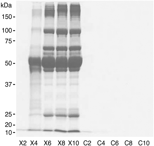
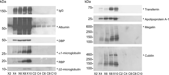
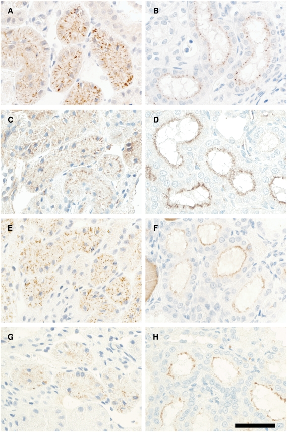
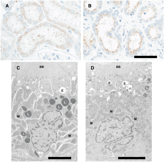
Methods: The investigation was performed in male XLAS dogs and age-matched normal male littermates. The urine profile and megalin-mediated endocytosis in the proximal tubule of six healthy and six XLAS dogs were examined at 2, 4, 6, 8 and 10 months of age using SDS-PAGE, immunoblotting and immunohistochemistry.

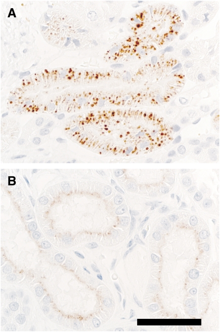
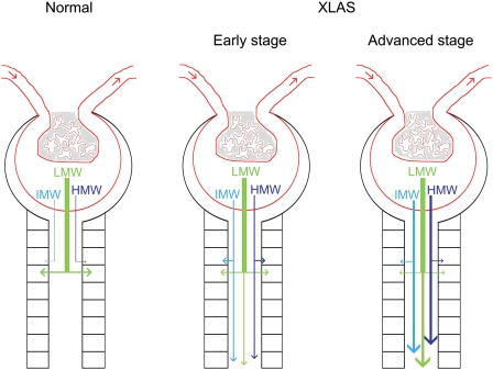
Results: Gradually increasing urinary excretion of proteins over time and a reduced content of the same proteins in proximal tubule cells were found. Besides the glomerular component of the proteinuria, a significant tubular component was seen, which is due to a progressive change in the uptake of low-molecular-weight (LMW) ligands by megalin. Furthermore, the protein overload present in the lumen of the proximal tubule exceeds the reabsorption capacity of megalin and the co-receptor cubilin and results in a combined low- and high-molecular-weight (HMW) proteinuria. Also, a shift in the distribution of lysosomes was seen in the XLAS dogs suggesting changes in the lysosomal degradation pattern in response to the altered endocytosis.

Conclusions: The present study shows that the increased glomerular permeability and the subsequently altered megalin-mediated and megalin-dependent cubilin-mediated endocytosis lead to a partial LMW proteinuria and partial HMW proteinuria.


Figures

Fig. 1
Fig. 1
SDS–PAGE gel showing a representative protein excretion pattern from a XLAS dog and a normal age-matched littermate. X2–X10 correspond to the serial urine samples from XLAS male dogs taken at 2, 4, 6, 8 and 10 months of age, respectively. C2–C10 correspond to the serial urine samples from normal male littermates also taken at 2, 4, 6, 8 and 10 months of age, respectively. The amounts of urine applied were normalized for creatinine concentration. Proteins were identified by Coomassie staining. Proteinuria is detectable in XLAS males at 4 months; urine protein levels appear to plateau at 6 months of age. No protein is found in control urine samples.
Fig. 2
Fig. 2
Immunoblotting of urine samples from XLAS and control male dogs showing differences in the urinary excretion of several megalin and/or cubilin ligands and the receptors megalin and cubilin. XLAS males exhibit progressively increasing levels of the ligands β2-microglobulin (MW, 12 kDa), retinol-binding protein (RBP) (MW, 22 kDa) and α1-microglobulin (MW, 26 kDa). The urinary levels of vitamin D binding protein (DBP) (MW, 56 kDa), albumin (MW, 66 kDa) and IgG (MW, 150 kDa) appeared to stagnate from the age of 4 months and for the rest of the measuring period. Urinary excretion of the solely cubilin ligands apolipoprotein A-1 (MW, 28 kDa) and transferrin (MW, 76 kDa) in XLAS dogs indicates impaired megalin-dependent cubilin-mediated endocytosis in addition to megalin-mediated endocytosis. Receptor fragment shedding of both receptors is present in the urine from XLAS dogs but virtually absent in the urine from the normal male littermates. X2–X10 correspond to the serial urine samples from XLAS male dogs taken at 2, 4, 6, 8 and 10 months of age, respectively. C2–C10 correspond to the serial urine samples from normal male littermates also taken at 2, 4, 6, 8 and 10 months of age, respectively. The amounts of urine applied were normalized for creatinine concentration. Primary antibodies used: a-α1-microglobulin 1:1500, a-retinol-binding protein (RBP) 1:1000, a-β2-microglobulin 1:400, a-vitamin D binding protein (DBP) 1:500, a-albumin 1:1000, a-IgG(H) 1:2000, a-apolipoprotein A-1 1:2000, a-transferrin 1:2000, a-megalin (GP330) 1:20 000 and a-cubilin (L403) 1:2000. HRP-conjugated secondary antibodies were used.
Fig. 3
Fig. 3
Reduced cellular content of the ligands α1-microglobulin (A, B), apolipoprotein A-1 (C, D), albumin (E, F) and IgG (G, H) was found in serial renal biopsies/necropsies from XLAS dogs by immunohistochemistry, indicating an impaired megalin-mediated and megalin-dependent cubilin-mediated endocytosis. The micrographs A, C, E and G are from the first biopsy taken at the age of 4 months, whereas the micrographs B, D, F and H are from the last biopsy/necropsy taken when the dogs were approximately 8 to 10 months old. There is a progressive reduced cellular content of all ligands, except albumin, which only show a reduced cellular content on the last tissue sample (necropsy). Primary antibodies used: a-α1-microglobulin 1:2400, a-apolipoprotein A-1 1:8000, a-albumin 1:100 000 and a-IgG(H) 1:800. HRP-conjugated secondary antibodies were used. Scale bar 50 μm.
Fig. 4
Fig. 4
Apical accumulation of proteins. At the age of 8–10 months, the ligands examined seem, despite a reduced reabsorbed amount, to be accumulating in the area just underneath the brush border of the proximal tubule cells in the XLAS dog (B) compared with the normal male littermate (A). Here, the labelling for the megalin ligand RBP is shown. Primary antibody used: a-RBP 1:10000. HRP-conjugated secondary antibodies were used. Scale bar 50 μm.
Fig. 5
Fig. 5
Change in lysosomal localization within proximal tubular epithelial cells. The distribution of lysosomes is shifted from the perinuclear compartment to the apical part of the proximal tubule cells in 8–10-months-old XLAS dogs compared with age-matched normal male littermates. Staining with a-lysosomal associated membrane glycoprotein 1 (LAMP-1) shows the change in lysosomal localization at the light microscopic level (A, B). Furthermore, as shown at the bottom of the figure, the localization of lysosomes (L) at electron microscopic level also seems to be shifted to the apical part of the proximal tubule cells (C, D). A and C are sections from normal male littermates and D are from XLAS male dogs. Primary antibody used: a-LAMP-1 1:1200. HRP-conjugated secondary antibody was used. Light microscopy: scale bar 50 μm. Electron microscopy: scale bar 5 μm. L, lysosomes; BB, brush border; E, endocytic vacuoles; M, mitochondria.
Fig. 6
Fig. 6
Composition of proteinuria during progression of the glomerular disease XLAS. Under normal physiologic conditions, LMW proteins are freely filtered and almost completely reabsorbed. There is little or no filtration of IMW and HMW proteins. In early stage of XLAS, filtered IMW and HMW proteins compete with LMW proteins for reabsorption, resulting in the appearance of LMW, IMW and HMW proteins in final urine. The uptake and degradation of protein by healthy proximal tubule cells remain intact. In the advanced stage of XLAS, uptake and degradation of filtered LMW, IMW and HMW proteins by damaged proximal tubule cells are abnormal, resulting in high levels of LMW, IMW and HMW proteins in final urine despite reduced GFR.

Similar articles

Cited by

References

    1. D'Amico G, Bazzi C. Pathophysiology of proteinuria. Kidney Int. 2003;63:809–825. - PubMed
    1. Maack T, Johnson V, Kau ST, et al. Renal filtration, transport, and metabolism of low-molecular-weight proteins: a review. Kidney Int. 1979;16:251–270. - PubMed
    1. Christensen EI, Nielsen R. Role of megalin and cubilin in renal physiology and pathophysiology. Rev Physiol Biochem Pharmacol. 2007;158:1–22. 1-22. - PubMed
    1. Christensen EI, Birn H. Multifunctional endocytic receptors. Nature reviews. 2002 - PubMed
    1. Birn H. The kidney in vitamin B12 and folate homeostasis: characterization of receptors for tubular uptake of vitamins and carrier proteins. Am J Physiol Renal Physiol. 2006;291:F22–F36. - PubMed

Publication types

MeSH terms

Substances