Review
. 2010 May;298(5):F1078-94.
doi: 10.1152/ajprenal.00017.2010. Epub 2010 Mar 3.

Acute kidney injury: a springboard for progression in chronic kidney disease

Affiliations
Review

Acute kidney injury: a springboard for progression in chronic kidney disease

Manjeri A Venkatachalam et al. Am J Physiol Renal Physiol. 2010 May.

Abstract

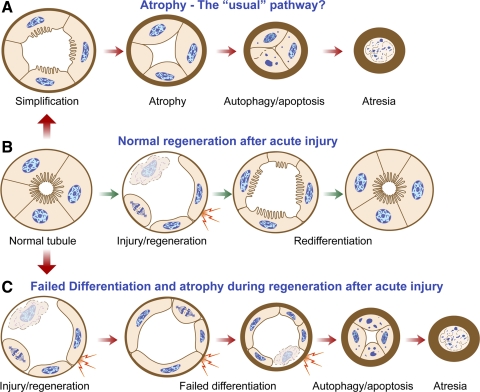
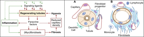
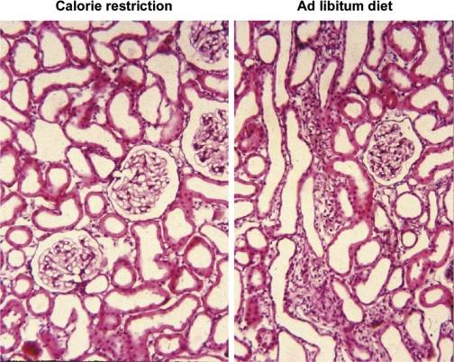
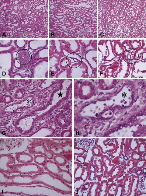
Recently published epidemiological and outcome analysis studies have brought to our attention the important role played by acute kidney injury (AKI) in the progression of chronic kidney disease (CKD) to end-stage renal disease (ESRD). AKI accelerates progression in patients with CKD; conversely, CKD predisposes patients to AKI. This research gives credence to older, well-thought-out wisdom that recovery from AKI is often not complete and is marked by residual structural damage. It also mirrors older experimental observations showing that unilateral nephrectomy, a surrogate for loss of nephrons by disease, compromises structural recovery and worsens tubulointerstitial fibrosis after ischemic AKI. Moreover, review of a substantial body of work on the relationships among reduced renal mass, hypertension, and pathology associated with these conditions suggests that impaired myogenic autoregulation of blood flow in the setting of hypertension, the arteriolosclerosis that results, and associated recurrent ischemic AKI in microscopic foci play important roles in the development of progressively increasing tubulointerstitial fibrosis. How nutrition, an additional factor that profoundly affects renal disease progression, influences these events needs reevaluation in light of information on the effects of calories vs. protein and animal vs. vegetable protein on injury and progression. Considerations based on published and emerging data suggest that a pathology that develops in regenerating tubules after AKI characterized by failure of differentiation and persistently high signaling activity is the proximate cause that drives downstream events in the interstitium: inflammation, capillary rarefaction, and fibroblast proliferation. In light of this information, we advance a comprehensive hypothesis regarding the pathophysiology of AKI as it relates to the progression of kidney disease. We discuss the implications of this pathophysiology for developing efficient therapeutic strategies to delay progression and avert ESRD.


Figures

Fig. 1.
Fig. 1.
Schematic diagram to illustrate how tubules regenerating after acute injury may fail to differentiate and exhibit profibrotic paracrine activity before they become atrophic. A: the “usual” pathway of tubule atrophy conceptualized from morphological studies of diseased kidneys involves simplification of epithelial structure progressing to autophagy and apoptosis accompanied by marked thickening of tubule basement membranes. Toward the end, tubules become atrectic and become enveloped by thick basement membranes or disappear altogether. B: normal regeneration of tubules after AKI with death of epithelium involves initial dedifferentiation, migration, and proliferation of surviving cells, followed by redifferentiation and full restoration of normal structure. Unlike in the usual pathway (A), tubule cells that survive acute kidney injury (AKI) follow a somewhat different route to become atrophic (C). Following dedifferentiation, migration, and proliferation, they fail to redifferentiate. After an indefinite period of time during which they are in a state of “failed differentiation,” these abnormal tubules proceed to develop thick basement membranes and undergo atrophy by autophagy and apoptosis just as in the usual pathway and disappear by atresia. Hyperactive epithelial paracrine signaling (shown as lightning bolts) in proliferating cells during regeneration becomes suppressed again if tubules redifferentiate normally (B) but persists in tubules with a regenerative failed differentiation phenotype (C) for an indefinite period of time before atrophy takes place. This paracrine signaling gives rise to inflammation and fibrosis, as depicted in Fig. 2.
Fig. 2.
Fig. 2.
Left: hypothetical scheme for relationships between signaling activity in regenerating tubule cells and downstream signaling events in the interstitium: inflammation, fibroblast proliferation, and capillary rarefaction. Although epithelial signaling with ensuing paracrine activity is the proximate “trigger” in this scheme, subsequent interactions between inflammation, fibroblasts, capillary endothelium, and tubule epithelium become a self-reinforcing “vicious cycle” that makes the separation of early signals and downstream events difficult. Right: pathology of tubulointerstitial fibrosis. Operation of the complex interactive signaling network depicted on the left leads to transformation of the normal tubulointerstitium (A) to a state of tubulointerstitial fibrosis: undifferentiated/atrophic tubules, proliferation of fibroblast progenitors, decreased capillary density, and inflammation (B).
Fig. 3.
Fig. 3.
Tubulointerstitial fibrosis precedes glomerulosclerosis in the remnant kidney after nephrectomy and is prevented by caloric restriction. Paraffin sections of remnant kidneys of rats 4 wk after nephrectomy by the infarction method, perfusion fixed with formaldehyde, and stained with hematoxylin and eosin. Rats were fed a 21% casein-based diet ad libitum (right) or pair fed a 35% casein-based diet at 60% of total food weight consumed by the ad libitum group, so that both groups consumed equal amounts of protein. The ad libitum group, but not the calorie-restricted group will develop end-stage renal disease after 5 mo (148). Original magnification: ×200. Unpublished archival material from Ref. .
Fig. 4.
Fig. 4.
Sections of hypertrophied kidneys from rats subjected to nephrectomy by right nephrectomy and either ligation of 2 of 3 branches of the left renal artery (A, D, G, and H) or surgical excision of both poles of the left kidney (B, E, and I) and sections of intact kidneys of spontaneously hypertensive stroke prone (SHR-SP) rat (C, F, and J). Kidneys were obtained ∼8 wk after nephrectomy or after a comparable period of observation from SHR-SP rats. For the rats from which these sections were obtained, the averaged systolic blood pressures measured by radio telemetry over a 4-wk period preceding death were (in mmHg) nephrectomy (infarction) 214; nephrectomy (excision) 133; and SHR-SP 214. At death, kidneys were perfused with Karnovsky's fixative. Paraffin-embedded sections were stained with hematoxylin and eosin. AC: micrographs to show presence or absence of tubulointerstitial fibrosis. DF: micrographs to show presence or absence of occlusive arteriolosclerosis (circled). GJ: micrographs to show presence or absence of tubules with regenerative epithelium (star) and acute injury with apoptotic cells (asterisk). Original magnifications: ×100 (AC); ×600 (DF); ×400 (G, I, and J). H: magnified view of tubule marked with small asterisk in G. Unpublished archival material from Bidani-Griffin laboratories was evaluated by M. Venkatachalam. This material is related to but not derived from published work that examined the effects of hypertension alone, renal ablation alone or renal ablation with hypertension on the development of glomerular pathology (19, 56, 59).
Fig. 5.
Fig. 5.
Sections from kidneys of rats microembolized through the left renal artery with 20- to 30-μm-diameter acrylic microspheres and examined 4 wk later. Serial paraffin sections of methyl Carnoy's fixed tissue were stained with Masson's trichrome (A) or immunohistochemically stained for vimentin (B), PDGF-B (C), or PDGF receptor (PDGFR)-β (D). Original magnification ×200. Reproduced from Ref. with kind permission from Dr. Akira Hishida, first department of Medicine, Hamamatsu University School of Medicine, Hamamatsu, Japan. Reproduced from Am J Pathol 158: 75–85, 2001; with permission from the American Society for Investigative Pathology.
Fig. 6.
Fig. 6.
Sections from kidneys of rats microembolized through the left renal artery with 20- to 30-μm-diameter acrylic microspheres and examined 4 wk later. Serial paraffin sections of methyl Carnoy's fixed tissue were stained with Masson's trichrome (A), periodic acid-Schiff (B), Phaseolus vulgaris agglutinin, a marker differentiated for proximal tubule cells (C), or immunohistochemically stained for vimentin (D), PDGF-B (E), and PDGFR-β (F). Original magnification ×400. All images show the same tubule cross section with adjacent interstitium in serial sections. Reproduced from Ref. with kind permission from Dr. Akira Hishida, first department of Medicine, Hamamatsu University School of Medicine, Hamamatsu, Japan. Reproduced from Am J Pathol 158: 75–85, 2001; with permission from the American Society for Investigative Pathology.
Fig. 7.
Fig. 7.
Sections of kidneys 14 days after right nephrectomy to remove 50% of renal mass and 45 min of ischemia of left kidney followed by reperfusion (right) or corresponding nonischemic nephrectomy controls (left). Paraffin sections of tissue perfusion fixed with periodic acid-lysine-paraformaldehyde were stained with hematoxylin and eosin (H&E; top) or immunohistochemically stained for the differentiation marker Na+-K+-ATPase (middle) or PDGF-B (red) and PDGFR-β (green; bottom). The images are from the outer stripe of the outer medulla, chief site of original ischemic damage. Top and middle: reproduced from Ref. with permission from the American Society for Investigative Pathology. Bottom: unpublished archival tissue from Ref. . Original magnifications: ×100 (top); ×200 (middle and bottom).

Similar articles

Cited by

References

    1. Abrams JR, Tapp DC, Venkatachalam MA. Dietary influence and pathologic changes. In: Progressive Nature of Renal Disease, edited by Mitch WE. New York: Churchill Livingstone, 1992, p. 133–148
    1. Abrass CK. Cellular lipid metabolism and the role of lipids in progressive renal disease. Am J Nephrol 24: 46–53, 2004 - PubMed
    1. Abrass CK. Overview: obesity: what does it have to do with kidney disease? J Am Soc Nephrol 15: 2768–2772, 2004 - PubMed
    1. Abu-Amarah I, Bidani AK, Hacioglu R, Williamson GA, Griffin KA. Differential effects of salt on renal hemodynamics and potential pressure transmission in stroke-prone and stroke-resistant spontaneously hypertensive rats. Am J Physiol Renal Physiol 289: F305–F313, 2005 - PubMed
    1. Al-Awqati Q, Preisig PA. Size does matter: will knockout of p21(WAF1/CIP1) save the kidney by limiting compensatory renal growth? Proc Natl Acad Sci USA 96: 10551–10553, 1999 - PMC - PubMed

Publication types