. 2010 May 7;285(19):14330-7.
doi: 10.1074/jbc.M109.090373. Epub 2010 Mar 3.

A slow, tight binding inhibitor of InhA, the enoyl-acyl carrier protein reductase from Mycobacterium tuberculosis

Affiliations

A slow, tight binding inhibitor of InhA, the enoyl-acyl carrier protein reductase from Mycobacterium tuberculosis

Sylvia R Luckner et al. J Biol Chem. .

Abstract

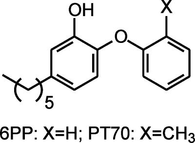
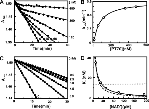
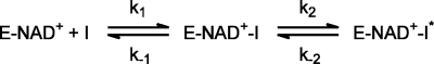
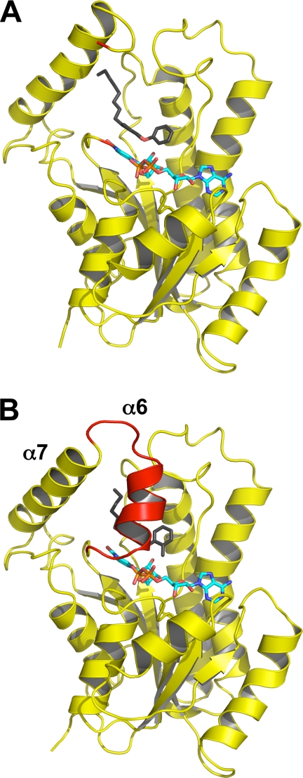
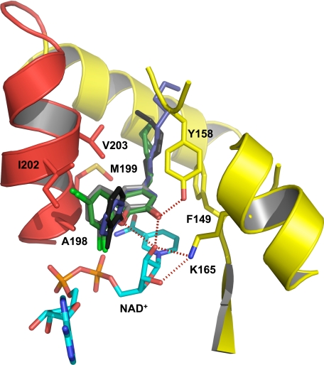
InhA, the enoyl-ACP reductase in Mycobacterium tuberculosis is an attractive target for the development of novel drugs against tuberculosis, a disease that kills more than two million people each year. InhA is the target of the current first line drug isoniazid for the treatment of tuberculosis infections. Compounds that directly target InhA and do not require activation by the mycobacterial catalase-peroxidase KatG are promising candidates for treating infections caused by isoniazid-resistant strains. Previously we reported the synthesis of several diphenyl ethers with nanomolar affinity for InhA. However, these compounds are rapid reversible inhibitors of the enzyme, and based on the knowledge that long drug target residence times are an important factor for in vivo drug activity, we set out to generate a slow onset inhibitor of InhA using structure-based drug design. 2-(o-Tolyloxy)-5-hexylphenol (PT70) is a slow, tight binding inhibitor of InhA with a K(1) value of 22 pm. PT70 binds preferentially to the InhA x NAD(+) complex and has a residence time of 24 min on the target, which is 14,000 times longer than that of the rapid reversible inhibitor from which it is derived. The 1.8 A crystal structure of the ternary complex between InhA, NAD(+), and PT70 reveals the molecular details of enzyme-inhibitor recognition and supports the hypothesis that slow onset inhibition is coupled to ordering of an active site loop, which leads to the closure of the substrate-binding pocket.


Figures

FIGURE 1.
FIGURE 1.
Structures of the rapid reversible inhibitor 6PP and the slow onset inhibitor PT70.
FIGURE 2.
FIGURE 2.
Progress curve analysis for the inhibition of InhA by PT70 and effect of NAD+ on the apparent inhibition constant of PT70. A, progress curves were obtained for inhibitor concentrations ranging from 0 to 480 nm. The solid curves are the best fits of the data to Equation 2. B, kobs plotted as a hyperbolic function of [PT70] using Equation 3. C, progress curves of InhA activity recovery were obtained for inhibitor concentrations ranging from 0 to 9 nm. The solid curves are the best fits of the data to Equation 2 to obtain kobs, which was then plotted against [PT70] using Equation 3 to obtain k−2. D, effect of NAD+ on the apparent inhibition constant of PT70. The fitted curves are shown for Equation 6 (dashed line; K1 = 0.0146 ± 0.0009 nm), Equation 7 (dotted line; K2 = 13.6 ± 5.5 nm), and Equation 8 (solid line; K1 = 0.022 ± 0.001 nm, K2 = 90.6 ± 9.7 nm).
FIGURE 3.
FIGURE 3.
Kinetic scheme for the slow onset inhibition of InhA by PT70.
FIGURE 4.
FIGURE 4.
Loop ordering upon slow binding inhibition. A, one monomer of the ternary InhA·NAD+·8PP complex (Protein Data Bank code 2b37) is shown in cartoon representation with the NAD+ molecule in cyan and the 8PP molecule in black and all-bonds representation. The substrate-binding loop is disordered in the 8PP structure, and the loop ends are depicted in red. B, monomer of the ternary InhA·NAD+·PT70 complex using the same colors and orientation as in A. The substrate-binding loop is ordered in this structure and covers the binding pocket (red cartoon). Secondary structure elements for both molecules were assigned with STRIDE (34).
FIGURE 5.
FIGURE 5.
Close-up of the binding pocket of InhA with bound NAD+ and PT70. Hydrogen bonds between PT70 (black) and Tyr158 (yellow) as well as the NAD+ molecule (cyan) are indicated as red dotted lines. The important hydrophobic residues Ala198, Met199, Ile202, and Val203 of the substrate-binding loop are shown in all bonds representation (red). An overlay of different triclosan inhibitors (PT70, black; triclosan, gray; 8PP, blue; JPL, green) in the binding pocket of InhA displays the differences in the orientation of the B-ring.

Similar articles

Cited by

References

    1. WHO (2007) WHO Report, WHO, Switzerland
    1. Jain A., Mondal R. (2008) FEMS Immunol. Med. Microbiol. 53, 145–150 - PubMed
    1. Zhang Y., Heym B., Allen B., Young D., Cole S. (1992) Nature 358, 591–593 - PubMed
    1. Banerjee A., Dubnau E., Quemard A., Balasubramanian V., Um K. S., Wilson T., Collins D., de Lisle G., Jacobs W. R., Jr. (1994) Science 263, 227–230 - PubMed
    1. Rawat R., Whitty A., Tonge P. J. (2003) Proc. Natl. Acad. Sci. U.S.A. 100, 13881–13886 - PMC - PubMed

Publication types

MeSH terms

LinkOut - more resources