. 2010 May 7;285(19):14424-37.
doi: 10.1074/jbc.M109.091108. Epub 2010 Mar 3.

NMR analysis of the structure, dynamics, and unique oligomerization properties of the chemokine CCL27

Affiliations

NMR analysis of the structure, dynamics, and unique oligomerization properties of the chemokine CCL27

Ariane L Jansma et al. J Biol Chem. .

Abstract

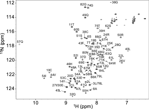
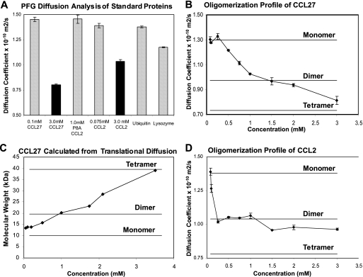
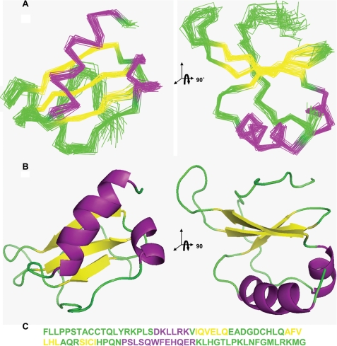
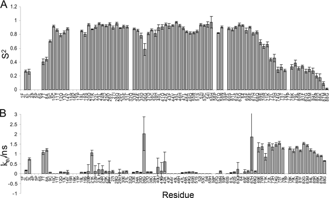
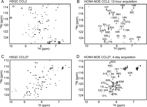
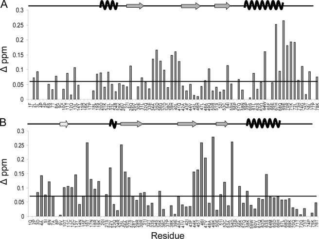
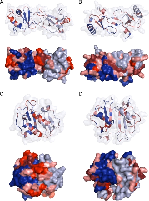
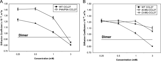
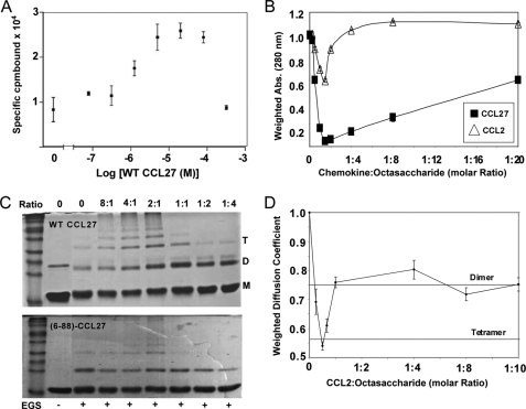
Chemokines have two essential interactions in vivo, with G protein-coupled receptors, which activate intracellular signaling pathways, and with glycosaminoglycans (GAGs), which are involved in cell surface localization and transport. Although it has been shown that chemokines bind and activate their respective G protein-coupled receptors as monomers, many chemokines oligomerize upon GAG binding, and the ability to oligomerize and bind GAGs is required for in vivo function. In this study, we investigated the structure, dynamics, and oligomerization behavior of cutaneous T-cell-attracting chemokine (CTACK, also known as CCL27) by NMR. (15)N relaxation and translational self-diffusion rates indicate that CCL27 oligomerizes, but in contrast to many other chemokines that form relatively discrete oligomers, CCL27 transitions between monomer, dimer, and tetramer species over a relatively narrow concentration range. A three-dimensional structure determination was pursued under conditions where CCL27 is primarily dimeric, revealing the standard motif for a chemokine monomer. Analysis of chemical shift perturbations of (1)H-(15)N HSQC spectra, relaxation-dispersion experiments, and filtered nuclear Overhauser effects suggest that CCL27 does not adopt a discrete CXC or CC dimer motif. Instead, CCL27 has uncommon oligomerization behavior, where several equilibria involving relatively low affinity interactions between different interfaces seem to be simultaneously at work. However, interaction with heparin avidly promotes oligomerization under conditions where CCL27 is monomeric by itself. We hypothesize that the plasticity in the oligomerization state may enable CCL27 to adopt different oligomeric structures, depending on the nature of the GAG binding partner, thereby providing a mechanism for increased diversity and specificity in GAG-binding and GAG-related functions.


Figures

FIGURE 1.
FIGURE 1.
Assigned 1H-15N HSQC of CCL27 acquired on a Bruker Avance II 600 MHz NMR with a 5-mm TCI CryoProbe. The sample was prepared at a concentration of 0.5 mm, in 50 mm acetate buffer, pH 5.6, at 302.3 K.
FIGURE 2.
FIGURE 2.
PFG diffusion analysis of CCL27. Assuming the diffusion coefficient at 0.075 mm corresponds to the monomeric form, the theoretical Ds value for the dimer was estimated using the Stokes-Einstein equation as 0.975 × 10⁻¹⁰ m2/s. A, PFG diffusion analysis of standard proteins with known molecular weights and oligomerization states. B, PFG diffusion analysis of CCL27 over a concentration range of 0.050 mm to 3.0 mm in 50 mm acetate, pH 5.6. C, molecular weight of CCL27 at different concentrations, calculated from the Ds values shown in B. D, PFG diffusion profile of WT CCL2 over a range of concentrations from 0.01 mm to 3.0 mm, in 50 mm acetate, pH 5.6.
FIGURE 3.
FIGURE 3.
Solution NMR structure of monomeric CCL27, displaying the standard chemokine structural motif: an N-loop (green) followed by a 310 helix (purple), three anti-parallel β-strands (yellow), and a C-terminal α-helix (purple). A, stereo views of the overlaid backbone traces for the 30 water-refined structures (residues Ala-8 to His-71). B, corresponding ribbon representation. C, sequence of CCL27 showing unstructured and loop regions in green, α-helices in purple, and β-strands in yellow.
FIGURE 4.
FIGURE 4.
Backbone 15N order parameters and internal correlation times from TENSOR2 analysis of relaxation data recorded on 1.0 mm CTACK at 500 MHz. Where the extended model-free formalism was required to fit the relaxation data, the order parameter shown is given by S2 = Ss2Sf2.
FIGURE 5.
FIGURE 5.
Filtered NOE analysis of a 50:50 15N-labeled plus 13C-labeled sample of CCL2 at 3.0 mm and a 50:50 15N-labeled (13C-depleted) plus 13C-labeled sample of CCL27 at 3.0 mm. A, 1H-15N HSQC of CCL2. B, two-dimensional 1H-15N (HC)NH-NOE, acquired in 12 h and assigned based on the HSQC. This experiment was previously employed to determine the dimeric structure of CCL2 in solution and used here to validate the method and compare with CCL27 (47). C, 1H-15N HSQC of CCL27. D, two-dimensional 1H-15N (HC)NH-NOE CCL27 at 3.0 mm, acquired in 4 days and assigned based on the HSQC.
FIGURE 6.
FIGURE 6.
1H-15N HSQC chemical shift perturbation analysis of CCL27 and CCL2. A, histogram of the change in chemical shift for residues 1–78 of CCL27, comparing data from 1.0 mm with 0.025 mm samples. Residues 79–88 displayed minimal shift changes and were excluded to directly compare CCL27 and CCL2 histograms. The monomeric secondary structural elements are displayed above. B, histogram of the change in chemical shift for each residue of CCL2, comparing data from 1.0 mm and 0.05 mm samples. The secondary structural elements are displayed above, including the N-terminal β-strand formed between the two monomeric subunits in the dimer structure (striped). The lines in both figures represent changes in chemical shift determined to be above baseline.
FIGURE 7.
FIGURE 7.
Results from chemical shift perturbation mapped to dimeric forms of CCL27 and CCL2, using a color gradient to depict the intensity of chemical shift change: 0.06–0.09 ppm in pink, 0.10–0.15 in maroon, and >0.16 in red. A, model of CCL27 forming a CC chemokine dimer. B, dimeric solution structure of CCL2. C, model of CCL27 forming a CXC chemokine dimer. D, the CXC dimer-like interface of the tetrameric form of CCL2.
FIGURE 8.
FIGURE 8.
PFG diffusion analysis of WT and mutant CCL27. A, results for mutations of the double N-terminal proline mutant, P4AP5A. B, PFG diffusion results for N-terminal truncations, resulting in monomeric variants of CCL27 based on theoretical calculations.
FIGURE 9.
FIGURE 9.
GAG binding analysis of CCL27 and CCL2. A, radioactive heparin-Sepharose binding assay of WT CCL27. The concentration of 125I-CTACK was fixed at 2.0 nm. Specifically bound 125I-CTACK was determined by subtracting Sepharose-bound from heparin-Sepharose-bound counts. B, solubility analysis of 0.1 mm CCL2 and CCL27 in the presence of increasing amounts of heparin octasaccharide. Solubility was determined by measuring the absorbance at 280 nm and normalizing to the value at 0 mm octasaccharide. C, chemical cross-linking of WT and the monomeric mutant (6–88)-CCL27 using Sulfo-EGS in the presence of increasing molar amounts of the GAG heparin decasaccharide. The molar ratios of CCL27:heparin decasaccharide are indicated above the gels and the presence of Sulfo-EGS is indicated below. D, 13C-edited PFG diffusion profile for 0.1 mm CCL2 with increasing concentration of heparin octasaccharide. Diffusion coefficients were normalized to the value at 0 mm octasaccharide, and the resulting ratios were also used to label the lines corresponding to the theoretical dimer and tetramer.

References

    1. Baggiolini M. (1998) Nature 392, 565–568 - PubMed
    1. Baggiolini M., Dewald B., Moser B. (1997) Annu. Rev. Immunol. 15, 675–705 - PubMed
    1. Charo I. F., Ransohoff R. M. (2006) N. Engl. J. Med. 354, 610–621 - PubMed
    1. Hartley O., Dorgham K., Perez-Bercoff D., Cerini F., Heimann A., Gaertner H., Offord R. E., Pancino G., Debré P., Gorochov G. (2003) J. Virol. 77, 6637–6644 - PMC - PubMed
    1. Kunkel E. J., Butcher E. C. (2002) Immunity 16, 1–4 - PubMed

Publication types

LinkOut - more resources