. 2010 Jun 25;285(26):19821-32.
doi: 10.1074/jbc.M110.104273. Epub 2010 Mar 3.

Stromal hyaluronan interaction with epithelial CD44 variants promotes prostate cancer invasiveness by augmenting expression and function of hepatocyte growth factor and androgen receptor

Affiliations

Stromal hyaluronan interaction with epithelial CD44 variants promotes prostate cancer invasiveness by augmenting expression and function of hepatocyte growth factor and androgen receptor

Shibnath Ghatak et al. J Biol Chem. .

Abstract

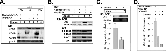
The main aim of our study is to determine the significance of the stromal microenvironment in the malignant behavior of prostate cancer. The stroma-derived growth factors/cytokines and hyaluronan act in autocrine/paracrine ways with their receptors, including receptor-tyrosine kinases and CD44 variants (CD44v), to potentiate and support tumor epithelial cell survival. Overexpression of hyaluronan, CD44v9 variants, and stroma-derived growth factors/cytokines are specific features in many cancers, including prostate cancer. Androgen/androgen receptor interaction has a critical role in regulating prostate cancer growth. Our previous study showed that 1) that increased synthesis of hyaluronan in normal epithelial cells promotes expression of CD44 variants; 2) hyaluronan interaction with CD44v6-v9 promotes activation of receptor-tyrosine kinase, which stimulates phosphatidylinositol 3-kinase-induced cell survival pathways; and 3) CD44v6/short hairpin RNA reduces colon tumor growth in vivo (Misra, S., Hascall, V. C., De Giovanni, C., Markwald, R. R., and Ghatak, S. (2009) J. Biol. Chem. 284, 12432-12446). Our results now show that hepatocyte growth factor synthesized by myofibroblasts associated with prostate cancer cells induces activation of HGF-receptor/cMet and stimulates hyaluronan/CD44v9 signaling. This, in turn, stabilizes the androgen receptor functions in prostate cancer cells. The stroma-derived HGF induces a lipid raft-associated signaling complex that contains CD44v9, cMet/phosphatidylinositol 3-kinase, HSP90 and androgen receptor. CD44v9/short hairpin RNA reverses the assembly of these components in the complex and inhibits androgen receptor function. Our results provide new insight into the hyaluronan/CD44v9-regulated androgen receptor function and the consequent malignant activities in prostate cancer cells. The present study describes a physiologically relevant in vitro model for studying the molecular mechanisms by which stroma-derived HGF and hyaluronan influence androgen receptor and CD44 functions in the secretory epithelia during prostate carcinogenesis.


Figures

FIGURE 1.
FIGURE 1.
Increased expression of HA promotes transformation of normal epithelial cells by augmenting expression and activation of the AR gene and soft agar colony formation. A–C, increased HA synthesis transforms PWR1E cells to be non-responsive to DT (10 ng/ml). HA and PSA in the medium were measured by an ELISA-like method (A) and the ELISA method (B), and colony formation in soft agar was detected by tetrazolium dye (C). The data in these experiments are representative of three experiments and, where indicated, are the mean of three experiments ± S.D. (error bars).
FIGURE 2.
FIGURE 2.
Increased HA stimulates CD44v9 expression, and silencing CD44v9 inhibits expression of AR and activation of cMet and IGF1R. Empty vector transfectant (lane 1) and untreated PWR1E-HAS2 (lane 2) were controls. PWR1E-HAS2 stable transfectants were transfected with control siRNA (lane 3), control shRNA (lane 4), CD44v9 siRNA (lane 5), or pSicoRCD44v9 shRNA (lane 6) plasmids (0.5 μg/5 × 105 cells). Expressions of proteins and message levels were determined by Western blots (WB) from the lysates and mRNA by RT-PCR from the total RNA of the transfectants. The data in these experiments are representative of three experiments.
FIGURE 3.
FIGURE 3.
Endogenous HA/CD44v9 interactions are required for AR gene expression, cell adhesion, and cell proliferation. A–C, an endogenous HA/CD44v9 interaction regulates AR-mediated gene expression. A, C4-2 cells were transfected with scrambled siRNA (control siRNA; lane 1), pSicoR scrambled shRNA (control shRNA; Control (1) (lane 2)), CD44v9 siRNA (lane 3), or pSicoR-CD44v9 shRNA (v9shRNA; lane 4) plasmids. PSA secreted in the media was measured by an ELISA method. Expressions of CD44v9 at protein and message levels were analyzed by Western blot (WB) and by RT-PCR. The lanes in RT-PCR are compared with lanes of Western blot and bars of PSA secretion against each treatment. B, HA/CD44 interaction is required for AR gene expression and cell proliferation. C4-2 (20 × 103) cells were transfected with control siRNA (lane 1), HAS2 siRNA (lane 2), pSicoR scrambled shRNA (Control (1), lane 3), or CD44v9 shRNA (lane 4). Cell proliferation was measured by an MTS assay and expressed as the mean absorbance at 490 nm/20 × 103 cells/90 min. PSA contents were measured by an ELISA. The lanes for cell proliferation of the upper panel are compared with lanes of PSA secretion against each treatment. C, CD44v9 regulates AR expression and function, whereas CD44v5 and CD44v10 do not. C4-2 cells were transfected with pSicoR scrambled shRNA (Control (1), lane 2), pRS scrambled shRNA (Control (2), lane 3), pRS-AR shRNA (lane 4), pSicoR-CD44v9 shRNA (lane 5), pSicoR-CD44v5 shRNA (v5shRNA) (lane 6), or pSicoR-CD44v10shRNA (v10shRNA) (lane 7). Non-transfected C4-2 cells were used as control (lane 1). The mRNAs for AR and glyceraldehyde-3-phosphate dehydrogenase mRNAs were measured by RT-PCR. In a separate experiment, the transcriptional activities of AR were measured, and the results are expressed as RLU/β-galactosidase activity. The lanes in RT-PCR are compared with bars of transcriptional activity of AR (RLU/β-galactosidase activity) against each of the plasmids. The data in these experiments are representative of three experiments and, where indicated, are the mean of three experiments ± S.D. (error bars).
FIGURE 4.
FIGURE 4.
AR regulates HA-mediated interaction of AR and CD44v9, cell invasion, and AR gene function. A, top, results of immunoprecipitation (IP) with AR followed by Western blot (WB) analysis of CD44 protein in the lysates of C4-2 treated with or without 10 ng/ml DT, 500 μg/ml HA for 24 h. A, bottom, DT/HA-treated and -untreated C4-2 cell (5 × 10⁴ cells/cm2) invasion in 24-well plates containing a Matrigel chamber on PCMFs (5 × 10⁴ cells/cm2). The lanes in the Western blot are compared with those of invasion against each of the treatments. B, C4-2 cells (1 × 10⁶), LNCaP cells (1 × 10⁶), or their transfectants with pSicoR scrambled shRNA (control shRNA) or pSicoR-CD44v9 shRNA (v9shRNA) plasmids were cultured in the 24-well tissue culture wells coated with supernatant from the respective cultures treated with 10 ng/ml DT or 10 ng/ml DT plus 500 μg/ml HA. Adherent cells were analyzed. Results are presented as percentage of adherence of non-transfected cells as control. C, the experiments in B were repeated for a fixed time period of 60 min. The data in this experiment are presented as control shRNA (lane 1), control shRNA plus HA (lane 2), and CD44v9 shRNA plus HA (lane 3). The cells were cultured for 48 h and then incubated with serum-free medium for 24 h. PSA secreted in the media was measured by ELISA. The data in these experiments are representative of three experiments and, where indicated, are the mean of three experiments ± S.D. (error bars).
FIGURE 5.
FIGURE 5.
Carcinoma cells induce HGF in PCMFs that stimulates HA production and HA/CD44v9-induced invasion. A, PCMFs (5 × 10⁴ cells/cm2) were co-cultured in CM from the C4-2 cells (5 × 10⁴ per/cm2) in the absence or presence of antibodies (10 μg/ml) to IL1R (lane 2), TNF-α (lane 3), IGF1 (lane 4), platelet-derived growth factor (lane 5), basic fibroblast growth factor (lane 6), or epithelial growth factor receptor (lane 7). HGF contents in the supernatants of the co-cultures were determined by ELISA. B, C4-2 cells (5 × 10⁴ cells/cm2) were grown in the absence and presence of control IgG or of 10 μg/ml anti-HGF Ab for 24 h, followed by treatment with 10 ng/ml HGF for another 48 h. CM were isolated from these cultures. PCMFs (5 × 10⁴ cells/cm2) were grown in the CM (in 1:1 (v/v) CM/SDM as described under “Experimental Procedures”) from untreated C4-2 cells (lane 1), CM from the C4-2 cells that were treated with HGF-antibody (lane 2), fresh SDM plus CM from IgG-treated C4-2 cells (lane 3), or fresh SDM plus CM from the HGF-antibody treated C4-2 cells (lane 4), followed by culture for 24 h. The concentrations of HA in the media of these CM co-cultures were determined by an ELISA-like method. C, top, CM from the C4-2 cells (5 × 10⁴ cells/cm2) pretreated with IgG (10 μg/ml) or with HGF-antibody (10 μg/ml) or transfected with control pSicoR scrshRNA or pSicoR-CD44v9 shRNA were prepared. PCMFs were grown in the CM from the C4-2 cell (in 1:1 (v/v) CM/SDM) and cultured for another 24 h: IgG treatment (lane 1), HGF antibody treatment (lane 2), control shRNA transfection (lane 3), and v9shRNA transfection (lane 4). HGF in the media was measured by an ELISA method, and the ratios of HGF/total DNA content (by measuring fluorescence of the Hoechst dye 33258) for the cells were plotted (y axis) for various treatments (x axis). C, bottom, PCMFs (in the well) were co-cultured with C4-2 cells (in the insert) treated with IgG (lane 1), transfected with pSicoR scrambled shRNA (lane 2), treated with anti-HGF antibody (lane 3) (10 μg/ml), or transfected with pSicoR-CD44v9 shRNA (v9shRNA) (lane 4) (5 × 10⁴ cells/200 μl) of the invasion chamber and were incubated for 3 days. Invasive cells that migrated to the underside of the membrane were counted. The data are presented as mean ± S.D. (error bars) of three experiments.
FIGURE 6.
FIGURE 6.
HGF induces cell proliferation/migration and HA production. A, 70% confluent C4-2 cells (2.5 × 10⁴) were grown in serum-depleted medium (see legend to Fig. 3) in the presence of various doses of HGF for 72 h, and cell proliferation was measured by counting viable cells in a trypan blue dye exclusion assay with a hemocytometer. B, the wells of a 24-well Matrigel invasion chamber contained 400 μl of SDM with various doses of HGF. Migration of C4-2 cells (2 × 10⁴ cells/200 μl of medium were added in the insert) to the lower side of the membrane were counted (12 different fields). C, C4-2 cells were treated with 10 ng/ml HGF for various times and were allowed to grow for another 24 h in SDM, and HA released in the medium was measured by an ELISA-like method. The data are presented as mean ± S.D. (error bars) of three experiments.
FIGURE 7.
FIGURE 7.
Down-regulation of CD44v9 inhibits PI3K (PI3K)/AKT activation, AR gene expression, and cell migration in C4-2 cells. A, C4-2 cells transfected with pSicoR scrshRNA (control shRNA) or pSicoR-CD44v9 shRNA (v9shRNA) were cultured with HGF (10 ng/ml). Cell lysates were extracted at the indicated time points and were analyzed by Western blot (WB) for p-AKT, AKT (data not shown), CD44, and β-actin. B, C4-2 cells transfected with control shRNA (lanes 1 and 3), or pSicoR-CD44v9 shRNA (lanes 2 and 4) were cultured to 70% confluence. After a 24-h incubation in SDM, cells were stimulated with HGF (10 ng/ml) for 12 h. Cells were then cultured for 72 h; cell lysates were prepared and analyzed for p-cMet, cMet, p-AKT, or AKT by Western blot; and mRNAs were determined for AR and glyceraldehyde-3-phosphate dehydrogenase by RT-PCR. Note that basal HGF secretion of C4-2 cells in this medium is <0.10 ng/ml. C, C4-2 cells were cultured without (lane 1) or with LY294002 (10 μm) (lane 2) for 12 h, followed by transfection with PSA-luciferase and pSV-β-galactosidase, and grown for 48 h. Cell extracts were prepared and used for luciferase and β-galactosidase assays, and results are expressed as RLU/β-galactosidase activity. D, C4-2 cells were treated with LY294002 (10 μm) for 12 h (lane 3). C4-2 cells were transfected with pRS scrshRNA (control shRNA; lane 1), or pRS-AR shRNA (ARshRNA; lane 2). The transfected cells (2 × 10⁴ cells/200 μl of medium) were added in the transwell inserts and allowed to migrate for 16 h at 37 °C. Migrated cell numbers were compared with that of untreated control cells and expressed as -fold increase. The data in these experiments are representative of three experiments and, where indicated, are the mean of three experiments ± S.D. (error bars).
FIGURE 8.
FIGURE 8.
Silencing CD44v9 inhibits assembly of p-cMet, AR, HSP90, P110α/PI3K, and CD44 into lipid raftlike structures. A, lipid raft fractions 2–5 of the gradients were isolated by sucrose density centrifugation from lysates of C4-2 cells that had been pretreated with HGF (10 ng/ml for 12 h) or were transfected with pSicoR scrambled shRNA (Control shRNA) or pSicoR-CD44v9 shRNA (v9shRNA) prior to treatment with HGF. Immunoprecipitates (IP) were prepared with antibodies against cMet and Western blotted (WB) with antibodies against cMet, p-cMet, and CD44 as well as for the p110α/PI3K, p85/PI3K, AR, and β-actin. B, lipid raft fractions (fractions 2–5) from the same cultures were immunoprecipitated with a human CD44 (HCAM) antibody and then Western blotted for human CD44v9 (∼140 kDa), CD44s (∼90 kDa), AR (∼65 kDa), and β-actin (∼55-kDa loading control). C (a–c), C4-2 cells untreated (lane 1) or transfected with scrambled siRNA (control-siRNA; lane 2), HAS2 siRNA (lane 3), pSicoR scrambled shRNA (Control shRNA, lane 4), pSicoR-CD44v9 shRNA (CD44v9 shRNA, lane 5), or methyl-β-cyclodextrin (lane 6). After 72 h of culture, components of signaling complex were dissociated by 5 mm methyl-β-cyclodextrin at 37 °C for 1 h. Most of the components (AR, CD44, p-cMet, etc.) after dissociation with methyl-β-cyclodextrin reside in the bottom fractions 6–8, whereas some p-cMet resides in fractions 2–5. Thus, fractions 2–8 were isolated and pooled and immunoprecipitated with cMet, and these immunoprecipitates were Western blotted for p-cMet (a and left bars in b) and AR (a and right bars in c). The bands in b and c were analyzed by densitometry. The data in all of these experiments are representative of three experiments and, where indicated, presented as mean ± S.D. (error bars) of three experiments.
FIGURE 9.
FIGURE 9.
Proposed model for the cross-talk between stromal HGF and tumor cell-derived HA/CD44v9/Met-induced signaling in prostate cancer cells. Cancer cells and stroma-derived fibroblasts influence each others' development. The extracellular domain of CD44 variants that contain the sequence encoded for v9 and its interaction with HA are required for the HGF-dependent activation of its receptor Met and its downstream anti-apoptotic malignant signaling involving PI3K/AKT and AR. HSP90 protects AR and AKT from degradation. Fibroblast (PCMF)-derived HGF and HA synthesized in response to HGF and cancer cell-derived AR, CD44v9, and cMet are involved in the malignant behavior in DT-independent prostate cancer cells.

Similar articles

Cited by

References

    1. Jemal A., Siegel R., Ward E., Hao Y., Xu J., Murray T., Thun M. J. (2008) CA Cancer J. Clin. 58, 71–96 - PubMed
    1. Grossmann M. E., Huang H., Tindall D. J. (2001) J. Natl. Cancer Inst. 93, 1687–1697 - PubMed
    1. Aaltomaa S., Lipponen P., Tammi R., Tammi M., Viitanen J., Kankkunen J. P., Kosma V. M. (2002) Urol. Int. 69, 266–272 - PubMed
    1. Lokeshwar V. B., Rubinowicz D., Schroeder G. L., Forgacs E., Minna J. D., Block N. L., Nadji M., Lokeshwar B. L. (2001) J. Biol. Chem. 276, 11922–11932 - PubMed
    1. Lipponen P., Aaltomaa S., Tammi R., Tammi M., Agren U., Kosma V. M. (2001) Eur. J. Cancer 37, 849–856 - PubMed

Publication types

MeSH terms