. 2010 Apr 15;21(8):1423-34.
doi: 10.1091/mbc.e09-10-0847. Epub 2010 Mar 3.

Protein kinase Czeta regulates Cdk5/p25 signaling during myogenesis

Affiliations

Protein kinase Czeta regulates Cdk5/p25 signaling during myogenesis

Aurélie de Thonel et al. Mol Biol Cell. .

Abstract

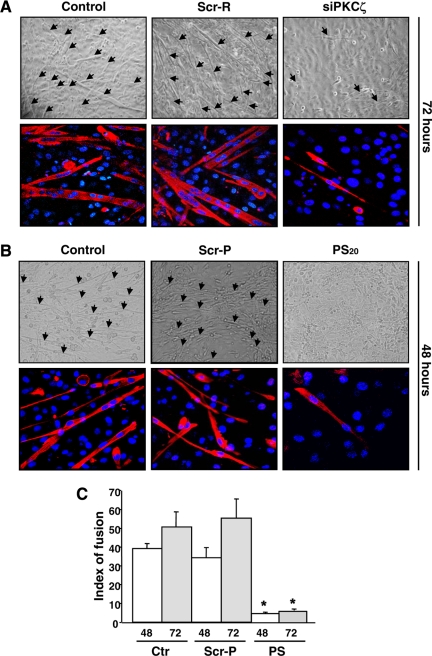
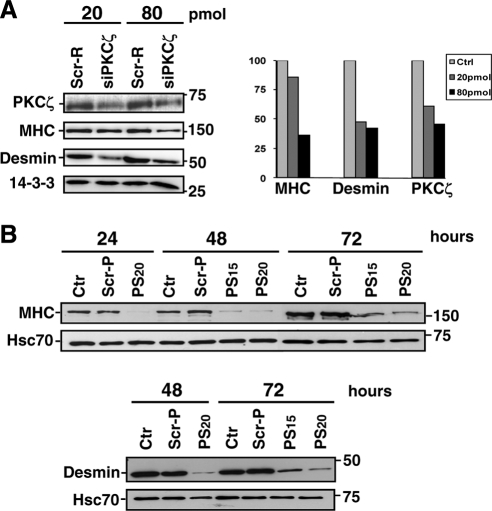
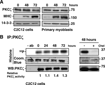
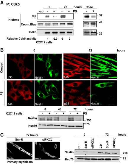
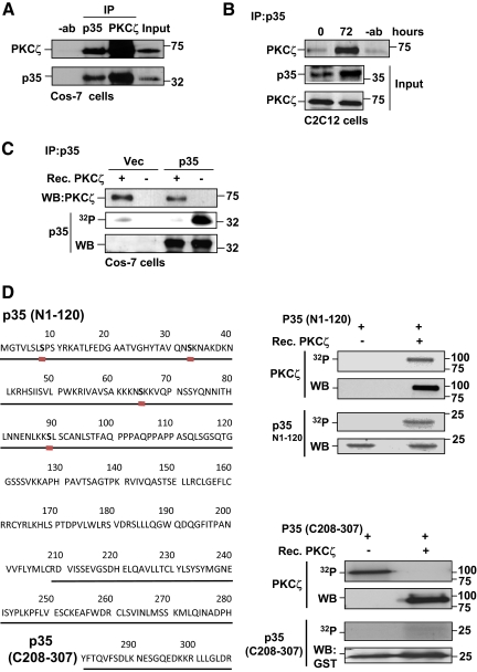
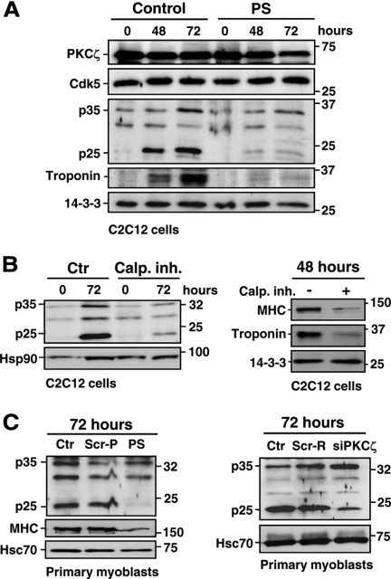
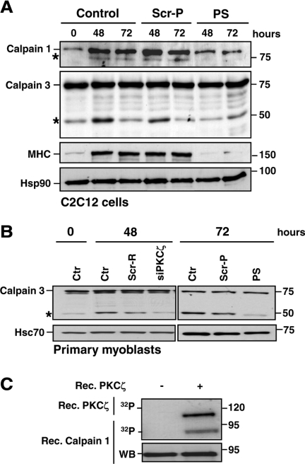
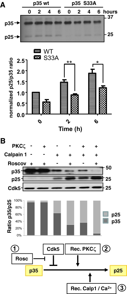
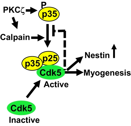
Atypical protein kinase Czeta (PKCzeta) is emerging as a mediator of differentiation. Here, we describe a novel role for PKCzeta in myogenic differentiation, demonstrating that PKCzeta activity is indispensable for differentiation of both C2C12 and mouse primary myoblasts. PKCzeta was found to be associated with and to regulate the Cdk5/p35 signaling complex, an essential factor for both neuronal and myogenic differentiation. Inhibition of PKCzeta activity prevented both myotube formation and simultaneous reorganization of the nestin intermediate filament cytoskeleton, which is known to be regulated by Cdk5 during myogenesis. p35, the Cdk5 activator, was shown to be a specific phosphorylation target of PKCzeta. PKCzeta-mediated phosphorylation of Ser-33 on p35 promoted calpain-mediated cleavage of p35 to its more active and stable fragment, p25. Strikingly, both calpain activation and the calpain-mediated cleavage of p35 were shown to be PKCzeta-dependent in differentiating myoblasts. Overall, our results identify PKCzeta as a controller of myogenic differentiation by its regulation of the phosphorylation-dependent and calpain-mediated p35 cleavage, which is crucial for the amplification of the Cdk5 activity that is required during differentiation.


Figures

Figure 1.
Figure 1.
Inhibition of PKCζ expression and activity prevents the formation of myotubes. (A) The down-regulation of PKCζ by siRNA abrogated the myoblast fusion and myotube formation. Primary myoblasts were transfected with PKCζ siRNA (siPKCζ) or scrambled siRNA (Scr-R) and induced to differentiate. At 72 h of differentiation, cells were fixed and double-stained with anti-MHC antibody, and DAPI, to detect differentiated cells and nuclei, respectively. The arrows in the phase-contrast images indicate myotubes. The bottom panels shows fluorescence labeling of MHC (red) and DNA (DAPI, blue), visualized by confocal microscopy. The images reveal a pronounced decrease in cell fusion and myotube formation as well as in MHC expression, when PKCζ is down-regulated. (B) The inhibition of PKCζ activity by a pseudosubstrate inhibitor peptide (PS) blocked the fusion of myoblast to multinucleated myotubes. Mouse primary myoblasts were induced to differentiate in the presence of either the PKCζ inhibitor peptide (PS; 15 or 20 μM) or the scrambled peptide (Scr-P; 20 μM). After 48 h of differentiation, cells were fixed and double-stained with antibodies against MHC and DAPI. Similarly to PKCζ down-regulation by siRNA (Figure 1A), PKCζ inhibition prevented myotube formation. (C) The fusion indexes of mouse primary myoblasts were determined in the presence of both the inhibitor peptide (PS, 20 μM) and the scrambled peptide (Scr-P, 20 μM). The fusion index was determined as a ratio of the number of nuclei in myotubes (showing at least two nuclei) to the total number of nuclei in six randomly chosen microscopic fields. The data are shown as means ± SEM (n = 3; *p < 0.05; Student's t test).
Figure 2.
Figure 2.
Inhibition of PKCζ expression and activity prevents myoblast differentiation. (A) The down-regulation of PKCζ by siRNA prevented the myoblast differentiation as indicated by the reduced amount of differentiation markers. Primary myoblasts were transfected with PKCζ siRNA (siPKCζ) or scrambled siRNA (Scr-R) and induced to differentiate. At the indicated time points of differentiation, cells were harvested for Western blot analysis. Desmin and MHC were used as markers of differentiation, whereas 14-3-3 served as a loading control. PKCζ down-regulation induced a decrease in expression of the differentiation markers MHC and desmin, as shown in the histogram. (B) The Inhibition of PKCζ activity by a pseudosubstrate inhibitor peptide (PS) blocked the myoblast differentiation. Mouse primary myoblasts were induced to differentiate in the presence of either the PKCζ inhibitor (PS; 15 or 20 μM) or the scrambled peptide (Scr-P; 20 μM). Cells were harvested at the indicated time points of differentiation and subjected to Western blot analysis. Increasing desmin and MHC expression levels indicated the progress of differentiation, whereas Hsc70 served as a loading control. Similarly to the PKCζ down-regulation by siRNA (Figure 2A), the inhibition of PKCζ activity prevented the myoblast differentiation.
Figure 3.
Figure 3.
Expression and activity of PKCζ during myogenesis. (A) The protein levels of PKCζ remained stable during myoblast differentiation. C2C12 cells and primary myoblasts were induced to differentiate, and cells were harvested at the indicated time points. The progress of differentiation was assessed by monitoring the expression of MHC, whereas 14-3-3 served as a loading control. (B) PKCζ activity was up-regulated during myoblast differentiation. C2C12 cells were shifted to differentiation medium, and PKCζ was immunoprecipitated from the cell lysates prepared at the indicated time points. The activity of immunoprecipitated PKCζ was analyzed using histone H1 as a substrate. As a negative control, cell extracts were incubated for 15 min with the PKC inhibitor chelerythrine (Chel, 5 μM) before the activity assay. Coomassie Blue staining was utilized to verify that equal amounts of substrate were used in each reaction, whereas immunoblot analysis confirmed that equal amounts of PKCζ were precipitated from each cell lysate. The blots were scanned on a densitometer to quantitate the amount of PKCζ and the degree of histone 1 phosphorylation. The ratio of 32P-histone 1 and PKCζ at each time point was calculated and normalized to the control values at time point 0 h. The data are representative of three independent experiments.
Figure 4.
Figure 4.
PKCζ modulates Cdk5 activity in vivo and affects the reorganization of nestin filaments during differentiation. (A) The Cdk5 activity was inhibited upon PKCζ inhibition. C2C12 cells were induced to differentiate for 72 h in the presence or absence of the PKCζ pseudosubstrate inhibitor peptide (PS). Cell lysates were subjected to the immunoprecipitation of endogenous Cdk5 and the Cdk5 kinase activity assays were performed using histone H1 as substrate (left). The numbers represent the relative induction in the Cdk5 activity during differentiation in the presence or absence of the inhibitor peptide PS. The data are representative of three independent experiments. As a negative control, cell lysates were preincubated with the Cdk5 inhibitor roscovitine (Rosc; 5 μM) for 15 min before kinase activity assay (right). Immunoblotting confirmed that equal amounts of Cdk5 were immunoprecipitated from each cell lysate. In addition, Coomassie Blue staining showed that equal amounts of histone 1 substrate were used in each reaction. (B) PKCζ activity was necessary for the reorganization of nestin filaments and the regulation of nestin protein levels during differentiation. C2C12 myoblasts were induced to differentiate for 72 h in the presence or absence of the inhibitor peptide (PS). The samples were fixed and double-stained with antibodies recognizing nestin and p35. At 72 h of differentiation, nestin filaments were aligned parallel to myotubes and colocalized with p35. However the Cdk5-mediated reorganization of nestin filaments during differentiation was disturbed when PKCζ activity was inhibited (scale = 10 μm, as in panel C). In parallel cell lysates were prepared from the same experiment, proteins were resolved by SDS-PAGE and analyzed by Western blotting for nestin and Hsc70, the latter serving as a loading control. The immunoblot revealed pronounced decreases in nestin protein levels when PKCζ was inhibited. (C) The down-regulation of PKCζ severely impaired nestin reorganization and stability during differentiation of primary myoblasts. Mouse primary myoblasts were transfected with PKCζ siRNA (siPKCζ) or scrambled siRNA (Scr-R) and induced to differentiate for 72 h. Samples were fixed, stained with anti-nestin antibody (scale bar, 10 μm), and analyzed by Western blotting. The results confirmed alterations in nestin remodeling and turnover observed during differentiation when PKCζ was down-regulated.
Figure 5.
Figure 5.
p35 interacts with and is a target for PKCζ. (A) PKCζ interacted with p35 in Cos-7 cells. Cos-7 cells were transfected with plasmids encoding p35 and PKCζ. Cells were lysed and subjected to immunoprecipitation using either anti-PKCζ or anti-p35 antibodies. Immunoprecipitated samples were separated on SDS-PAGE together with input controls and immunoblotted for PKCζ and p35. The results revealed an interaction between p35 and PKCζ. (B) An interaction between p35 and PKCζ was detected in differentiating myoblasts. C2C12 cells were induced to differentiate, lysed, and subjected to p35 immunoprecipitation. Western blot analysis of immunoprecipitates indicated that p35 and PKCζ interacted during differentiation. (C) p35 was phosphorylated by PKCζ. Cos-7 cells were transfected with p35 or an empty vector. 48 h later, cells were lysed, and p35 was immunoprecipitated. An in vitro phosphorylation assay with recombinant PKCζ was performed using immunoprecipitated p35 as a substrate. The immunoblots demonstrate the amounts of p35 and PKCζ and the autoradiograph the degree of phosphorylation in the different reactions. (D) The N-terminus of p35 contains several putative PKCζ phosphorylation sites (the ProteinScan program was helpful in the prediction of putative phosphorylation sites; see http://156.40.231.198/ProteinScan/ScanProteinForPKCSitesPage.aspx). The N-terminus of p35 was specifically phosphorylated in vitro by PKCζ. In vitro phosphorylation assays were performed with recombinant PKCζ using either truncated N-terminal (N1-120) or C-terminal (C208-307) p35 peptide as a substrate. Proteins were resolved by SDS-PAGE, and the presence of indicated proteins in phosphorylation reactions was monitored by Western blotting. Autoradiographs demonstrate the phosphorylation of p35 peptides as well as the autophosphorylation of PKCζ (indicating kinase activity). Remarkably, only the N-terminus of p35 (N1-120) was specifically phosphorylated by PKCζ.
Figure 6.
Figure 6.
PKCζ activity regulates the calpain-mediated cleavage of p35. (A) The calpain-mediated cleavage of p35 during differentiation was prevented by the PKCζ inhibition. C2C12 cells were induced to differentiate in the presence or absence of the pseudosubstrate inhibitor peptide (PS; 20 μM). Cells were harvested at the indicated time points and the corresponding lysates were subjected to immunoblotting using the indicated antibodies. The immunoblot reveals that p35 is cleaved to p25 fragment during the differentiation and that this cleavage is inhibited in the presence of the inhibitor peptide PS. (B) The inhibition of calpain activity by the calpain inhibitor III prevented the p25 generation and inhibited the differentiation of C2C12 myoblasts. C2C12 cells were induced to differentiate in the presence of the calpain inhibitor III (15 μM) and harvested at the indicated time points. Cell lysates were analyzed by Western blotting using the indicated antibodies. (C) The inhibition of PKCζ activity and expression prevents the p35 processing to p25. Primary myoblasts were induced to differentiate in the presence or absence of the inhibitor peptide (PS) or a scrambled peptide (Scr-P; 20 μM; left), or, alternatively, after the transfection with PKCζ siRNA (siPKCζ) or scrambled siRNA (Scr-R; 60 pmol; right). Cell lysates were immunoblotted with the indicated antibodies. Troponin and MHC were used as markers of differentiation, whereas Hsc70, Hsp90, and 14-3-3 served as loading controls.
Figure 7.
Figure 7.
PKCζ regulates the calpain activity during differentiation. (A) PKCζ depletion inhibited the activation of calpains. C2C12 cells were induced to differentiate in the presence of either the pseudosubstrate inhibitor peptide (PS) or scrambled peptide (Scr-P; 20 μM). Samples were harvested at the indicated time points and analyzed by immunoblotting using the indicated antibodies. The activation of calpains during differentiation was monitored by the formation of the auto-cleavage products (denoted by asterisk) of calpain 1 and the muscle-specific isoform, calpain 3. MHC was used as a marker of differentiation, whereas Hsp90 served as a loading control. The immunoblots indicate significant reduction in calpain activation during differentiation when PKCζ activity was inhibited. (B) Mouse primary myoblasts were induced to differentiate after transfection with PKCζ siRNA (siPKCζ) or scrambled siRNA (Scr-R) or in the presence of the pseudosubstrate inhibitor peptide (PS) or a scrambled peptide (Scr-P). Samples were harvested at the indicated time points and subjected to immunoblotting using the indicated antibodies. Hsc70 served as a loading control. The results verified the observations presented in A establishing PKCζ as a regulator of calpains. (C) Calpain 1 is phosphorylated in vitro by PKCζ. A phosphorylation assay was performed with recombinant PKCζ using recombinant calpain 1 as a substrate. Proteins were separated by SDS-PAGE and transferred to nitrocellulose membranes. First, the phosphorylation of calpain 1 as well as the PKCζ autophosphorylation, indicating kinase activity, were detected by autoradiography and, second, the membranes were subjected to Western blot analysis.
Figure 8.
Figure 8.
p35 phosphorylation by PKCζ regulates the calpain-mediated cleavage of p35. (A) PKCζ-induced serine 33 phosphorylation of p35 promotes the p35 cleavage. Site-directed mutagenesis was used to replace serine 33 with alanine residue in p35. Plasmids encoding the WT and the S33A-mutated p35 were transfected to C2C12 myoblasts. To activate calpains in proliferating, nondifferentiating C2C12 myoblasts the calcium ionophore A23187 was utilized for the indicated time periods. The generation of p25 was assessed by Western blot analysis. The quantitative data represents mean values of three independent experiments. Mutation of serine 33 resulted in a decrease in the p25 formation indicating that the PKCζ-mediated phosphorylation promotes the calpain-mediated cleavage of p35. (B) PKCζ facilitated the calpain 1–mediated cleavage of p35 and overcame the inhibitory effect of Cdk5. An in vitro assay was conducted to investigate the effect of PKCζ- or Cdk5-mediated phosphorylation on the calpain dependent cleavage of p35. First a recombinant GST-p35/His-Cdk5 complex was incubated either 1) alone (lane 3), 2) with roscovitine (4 μM; lanes 1 and 4) to inhibit the Cdk5-dependent p35 phosphorylation, 3) with recombinant PKCζ (lanes 2 and 5), or 4) with both roscovitine and PKCζ (lane 6). Subsequently, samples were incubated with or without recombinant calpain 1 for 5 min at RT to induce the cleavage of p35. Samples were separated by SDS-PAGE and immunoblotted using anti-p35 antibody. The Cdk5 immunoblot served as a loading control. The quantitative analysis demonstrates the p35-p25 ratios. The results show that calpain 1 treatment alone did not produce as much p25 (lane 3) as when the Cdk5/p35 complex was prephosphorylated with PKCζ (lane 5). The inhibition of Cdk5 activity promoted the generation of p25 depicting the autoinhibitory effect stated in the literature (lane 4; Kamei et al., 2007). Interestingly, PKCζ was insensitive to the Cdk5-induced inhibition and largely increased the calpain-mediated p25 formation. The calpain-dependent generation of the p25 fragment was maximal in the presence of PKCζ and of the Cdk5 inhibitor roscovitine (lane 6). The first two lanes serve as a controls, proving that p25 is not generated in the absence of calpains. The rationale of how the PKCζ-controlled signaling complex would work, as judged from the presented results of the three different treatments and their combinations, is outlined in the scheme below the bottom panel.
Figure 9.
Figure 9.
A tentative model for PKCζ signaling during muscle differentiation. The up-regulation of PKCζ activity during myoblast differentiation promotes the activation of Cdk5 in a calpain-dependent manner. PKCζ phosphorylates the Cdk5 activator protein, p35, and calpains. Consecutively, calpains are activated and trigger the cleavage of the phosphorylated p35 to its more stable fragment, p25, leading to a sustained activation of Cdk5. PKCζ is able to break the autoinhibitory loop, where Cdk5 restrains the p35 processing and p25 generation by phosphorylating p35. Thus, PKCζ is a major upstream regulator of Cdk5 kinase activity essential for the progression of the myogenic differentiation.

Similar articles

Cited by

References

    1. Barnoy S., Glaser T., Kosower N. S. The calpain-calpastatin system and protein degradation in fusing myoblasts. Biochim. Biophys. Acta. 1998;1402:52–60. - PubMed
    1. Barnoy S., Maki M., Kosower N. S. Overexpression of calpastatin inhibits L8 myoblast fusion. Biochem. Biophys. Res. Commun. 2005;332:697–701. - PubMed
    1. Berra E., Diaz-Meco M. T., Dominguez I., Municio M. M., Sanz L., Lozano J., Chapkin R. S., Moscat J. Protein kinase C zeta isoform is critical for mitogenic signal transduction. Cell. 1993;74:555–563. - PubMed
    1. Castellani L., Salvati E., Alema S., Falcone G. Fine regulation of RhoA and Rock is required for skeletal muscle differentiation. J. Biol. Chem. 2006;281:15249–15257. - PubMed
    1. Cheung Z. H., Ip N. Y. Cdk5, mediator of neuronal death and survival. Neurosci. Lett. 2004;361:47–51. [Review] - PubMed

Publication types

MeSH terms

LinkOut - more resources