. 2010 May 1;21(9):1505-18.
doi: 10.1091/mbc.e09-06-0485. Epub 2010 Mar 3.

A myosin IK-Abp1-PakB circuit acts as a switch to regulate phagocytosis efficiency

Affiliations

A myosin IK-Abp1-PakB circuit acts as a switch to regulate phagocytosis efficiency

Régis Dieckmann et al. Mol Biol Cell. .

Abstract

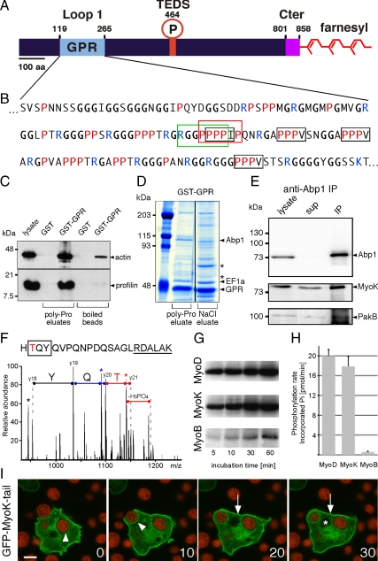
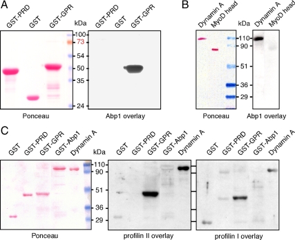
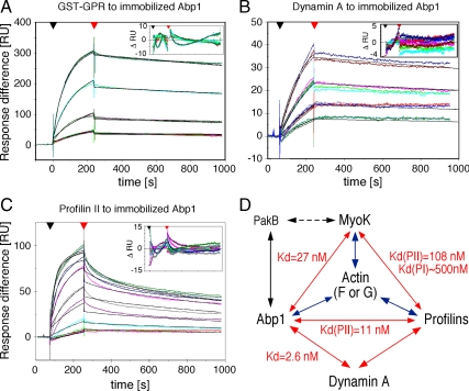
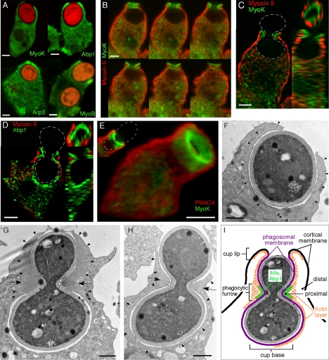
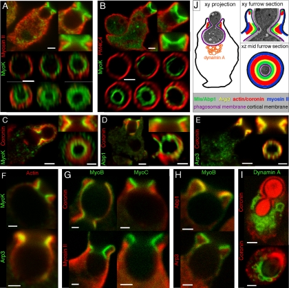
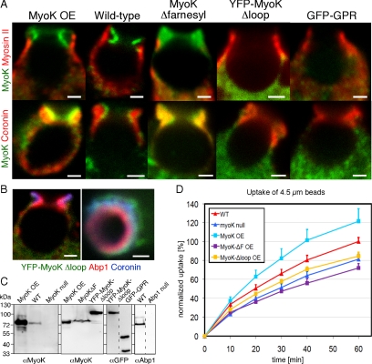
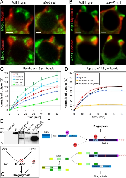
Actin dynamics and myosin (Myo) contractile forces are necessary for formation and closure of the phagocytic cup. In Dictyostelium, the actin-binding protein Abp1 and myosin IK are enriched in the closing cup and especially at an actin-dense constriction furrow formed around the neck of engulfed budded yeasts. This phagocytic furrow consists of concentric overlapping rings of MyoK, Abp1, Arp3, coronin, and myosin II, following an order strikingly reminiscent of the overall organization of the lamellipodium of migrating cells. Mutation analyses of MyoK revealed that both a C-terminal farnesylation membrane anchor and a Gly-Pro-Arg domain that interacts with profilin and Abp1 were necessary for proper localization in the furrow and efficient phagocytosis. Consequently, we measured the binding affinities of these interactions and unraveled further interactions with profilins, dynamin A, and PakB. Due to the redundancy of the interaction network, we hypothesize that MyoK and Abp1 are restricted to regulatory roles and might affect the dynamic of cup progression. Indeed, phagocytic uptake was regulated antagonistically by MyoK and Abp1. MyoK is phosphorylated by PakB and positively regulates phagocytosis, whereas binding of Abp1 negatively regulates PakB and MyoK. We conclude that a MyoK-Abp1-PakB circuit acts as a switch regulating phagocytosis efficiency of large particles.


Figures

Figure 1.
Figure 1.
The atypical domains of MyoK are functional. (A) MyoK contains a unique GPR domain (aa 122-265; pale blue) inserted in loop 1 of the motor domain. No neck, no light chain binding site, and no polybasic stretch can be recognized after the last conserved amino acids of the motor domain (KIF; aa 806-808). Instead, the atypical short tail (magenta) ends with a CAAX box (CLIQ; aa 855-858). (B) In the sequence of the GPR loop, the type I SH3 binding motif is boxed in green. The putative profilin binding poly-proline stretch is boxed in red. Unconventional profilin binding motifs, ZPPϕ (where Z is generally proline, glycine, or alanine and ϕ a hydrophobic residue) are boxed in black. (C) GST and GST-GPR coupled beads were incubated with wild-type cytosol and eluted with 0.1 mM PLP, showing that profilin-actin quantitatively dissociated from GST-GPR but not GST. (D) GST-GPR coupled beads were incubated with wild-type cytosol and eluted with 100 μM PLP and 1 M NaCl. Eluates were electrophoresed and gels Coomassie stained. Asterisks indicate two major unidentified protein bands. (E) MyoK and PakB were coimmunoprecipitated with anti-Abp1 antibodies. (F) MyoK is phosphorylated in vivo at the TEDS site, as revealed by the fragmentation spectrum of the peptide HTQYQVPQNPDQSAGLRDALAK that displays the y18-y21 sequence ions (2+) that identify the N-terminal TYQ amino acids (box) together with a neutral loss of H3PO4, characteristic of phosphorylated threonine. Peaks corresponding to H2O loss are indicated by stars. The phosphorylated threonine is in red and conserved residues neighboring the TEDS site are underlined. (G and H) MyoK, MyoD but not MyoB are phosphorylated in vitro by PakB. (G) The autoradiograms show the incorporation of 32P into purified myosin head constructs at indicated times of incubation with [γ-32P]ATP and purified GST-PakB. (H) The rates of phosphate incorporation were linear up to 60 min for all reactions. Data are representative of three independent assays. (I) The GFP-MyoK tail construct localized to the plasma membrane (arrow). It was not excluded from the phagocytic cup but progressively lost during maturation (arrowheads). The nucleus is indicated by an asterisk. Bar, 5 μm.
Figure 2.
Figure 2.
A network of direct protein-protein interactions linking the GPR loop of MyoK, Abp1, profilins, and dynamin A was revealed by blot overlays. GST-Abp1 directly interacts with immobilized GPR loop (A) and dynamin A (B). Purified profilin I and II were overlaid on membranes blotted in parallel (C). Both isoforms clearly interact with immobilized Abp1 and dynamin A. Weak interaction of profilin II with Abp1 and profilin I with WASp PRD are detected although the signal intensity of the control (GST) is similar. A Ponceau staining is shown (left) and corresponding overlays are shown (right). GST and the MyoD head domain were used as negative controls for GST fusion or His-tagged proteins, respectively.
Figure 3.
Figure 3.
SPR confirms and quantifies the direct interactions detected by blot overlays. (A–C) The affinity of the GPR loop (A), dynamin A (B), and profilin II (C) to immobilized GST-Abp1 were determined by SPR. A GST-coupled chip was used in the reference chamber. Experimental curves are in color, and fitted curves are in black. The range of concentrations of analytes used is indicated in Table 1. (D) Schematic representation of the protein-protein interaction network unraveled in this study. Arrows indicate direct interactions. When determined, equilibrium constants are indicated. Interactions with actin have been shown previously (blue arrows). A dashed arrow indicates indirect interaction identified by anti-Abp1 immunoprecipitation.
Figure 4.
Figure 4.
Enrichment of cytoskeletal proteins in the phagocytic cup and phagocytic furrow. (A) MyoK, Abp1, Arp3, and MyoB are enriched at the lip of the phagocytic cup. Cells were fed with TRITC-labeled fluorescent yeasts for 5 min. MyoK, Abp1, and Arp3 were localized by specific antibody detection in MyoK-overexpressing cells (MyoK OE) or wild-type cells, respectively. The GFP-MyoB signal was enhanced by anti-GFP antibodies. (B–E) Enrichment of MyoK and Abp1 around the bud neck of engulfed yeast defines the phagocytic furrow. Dotted white lines represent the contour of budded yeasts. (B) Serial averaged projections of three confocal sections of cells immunostained for MyoK and myosin II. (C) xy, yz, and xz plane sections of the series shown in B. (D) xy, yz, and xz plane sections of a cell immunostained for Abp1 and myosin II. (E) 3D reconstruction of a phagocytic furrow stained for MyoK and PM4C4. A single section of the same image is presented (top left corner). (F–H) Electron micrographs of phagocytosed yeasts in wild-type cells. (F) Phagocytic cup enclosing single yeast displayed actin enrichment not only around the phagosomal membrane (closed arrowheads) but also lining the cortical plasma membrane near the lip (open arrowheads). (G and H) Phagocytic cups enclosing budded yeasts at two stages of closure. Arrows point at the actin-rich zone of the phagocytic furrow. A denser dark area in the actin-rich zone of the furrow (open arrowheads) is particularly visible in G. The actin layer surrounding the cup is continuous with the actin-rich zone in the furrow (closed arrowheads). This actin layer is visible at the lip in G and H, whereas it is absent from the cup base in G. (I) Schematic representation of the phagocytic cup with a furrow and definition of the terms used in this study. Bars, 2 μm (A–E) or 1 μm (F–H).
Figure 5.
Figure 5.
Actin-binding proteins are enriched in distinct territories in the phagocytic furrow. (A–I) Sections (xy plane) and magnifications of the phagocytic furrow represent averaged projections over two or three confocal sections (0.3–0.5 μm depth). (A–F) Four to five distinct protein layers or territories are observed in sections through the furrow. A MyoK layer is surrounded by a myosin II (A) and plasma membrane (4C4) layer (B). Whereas there is a weak overlap with MyoK (C–F) and Abp1 (D), actin or coronin extensively overlap with Arp3 (E–F). For en face views (xz plane) through the phagocytic furrow, images were deconvoluted and reconstructed in 3D. The plane of the furrow at minimal ring diameter determined the midplane. A gallery of successive sections through the furrow, parallel to the midplane is also shown. (G–I) Confocal images illustrating the positioning in the phagocytic furrow of GFP-MyoB, YFP-MyoC, and GFP-dynamin A relative to the cytoskeletal proteins mentioned above. (J) Schematic representation of the different territories observed. For whole cells sections (A–E and I), bars are 2 μm. For magnified views of the furrow, bars are 1 μm.
Figure 6.
Figure 6.
The GPR loop and farnesylation motif are necessary for the fine localization of MyoK in the phagocytic furrow and optimal uptake of large particles. (A) Localization of MyoK relative to myosin II and coronin was monitored. MyoK was detected by antibody labeling in wild-type MyoK overexpresser (OE) and MyoKΔfarnesyl-expressing cells or expression of fluorescent fusion proteins YFP-MyoKΔloop and GFP-GPR. (B) Exclusion of cytosolic YFP-MyoKΔloop from the territory stained by Abp1 but not by coronin. Bars, 1 μm. (C) Immunoblots showing the expression levels of the MyoK constructs relative to the endogenous protein. (D) Flow cytometry uptake assay of 4.5-μm fluorescent latex beads of cells overexpressing MyoK full-length, MyoKΔloop, and MyoKΔfarnesyl proteins relative to MyoK null and wild-type parent strains. Data are expressed as a percentage of beads ingested by wild-type cells at 60 min.
Figure 7.
Figure 7.
Common and independent roles of MyoK and Abp1 in phagocytosis. (A) MyoK localized at the furrow and did not overlap with myosin II or coronin in wild-type or abp1 null cells, similarly to the overexpressed protein. (B) Localization and enrichment of Abp1 in the furrow was similar to MyoK and did not differ in wild-type or myoK null cells (bars, 2 μm). (C and D) The uptake of 4.5-μm fluorescent latex beads was quantitated by flow cytometry in myoK null cells, in abp1 null cells and in complemented cell lines overexpressing MyoK or Abp1, respectively (C). Uptake of beads was also measured in wild-type or myoK null cells overexpressing or not constitutively active PakBΔPL (D). Data are expressed as percentage of beads ingested by wild-type cells at 60 min, where 100% corresponds to 2.2 beads/cell. (E) Expression levels of endogenous PakB (top band) and overexpressed PakBΔPL (bottom band) in wild-type or myoK null cells. (F) Schematic representation of the regulatory protein–protein interactions responsible for circuit function. (G) Integrated model of the Abp1-MyoK-PakB circuit proposed to act as a switch regulating phagocytosis of big particles.

Similar articles

Cited by

References

    1. Bertling E., Quintero-Monzon O., Mattila P. K., Goode B. L., Lappalainen P. Mechanism and biological role of profilin-Srv2/CAP interaction. J. Cell Sci. 2007;120:1225–1234. - PubMed
    1. Cai L., Makhov A. M., Schafer D. A., Bear J. E. Coronin 1B antagonizes cortactin and remodels Arp2/3-containing actin branches in lamellipodia. Cell. 2008;134:828–842. - PMC - PubMed
    1. Clarke M., Maddera L. Phagocyte meets prey: uptake, internalization, and killing of bacteria by Dictyostelium amoebae. Eur. J. Cell Biol. 2006;85:1001–1010. - PubMed
    1. Crawley S. W., de la Roche M. A., Lee S. F., Li Z., Chitayat S., Smith S. P., Cote G. P. Identification and characterization of an 8-kDa light chain associated with Dictyostelium discoideum MyoB, a class I myosin. J. Biol. Chem. 2006;281:6307–6315. - PubMed
    1. Dai J., Ting-Beall H. P., Hochmuth R. M., Sheetz M. P., Titus M. A. Myosin I contributes to the generation of resting cortical tension. Biophys. J. 1999;77:1168–1176. - PMC - PubMed

Publication types

MeSH terms

LinkOut - more resources