Review
. 2010 Mar 15;123(Pt 6):825-35.
doi: 10.1242/jcs.064790.

Welcome to a new kind of tension: translating kinetochore mechanics into a wait-anaphase signal

Affiliations
Review

Welcome to a new kind of tension: translating kinetochore mechanics into a wait-anaphase signal

Thomas J Maresca et al. J Cell Sci. .

Abstract

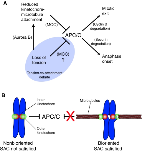
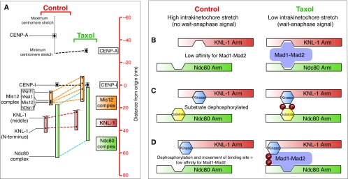
Recent high-resolution studies of kinetochore structure have transformed the way researchers think about this crucial macro-molecular complex, which is essential for ensuring chromosome segregation occurs faithfully during cell division. Kinetochores mediate the interaction between chromosomes and the plus-ends of dynamic spindle microtubules and control the timing of anaphase onset by regulating the spindle assembly checkpoint (SAC). There is much debate in the SAC research community as to whether mitotic cells sense only microtubule attachment at the kinetochore, or both attachment and tension, before committing to anaphase. In this Commentary, we present a brief history of the tension-versus-attachment debate, summarize recent advances in our understanding of kinetochore structure and focus on the implications of a phenomenon known as intrakinetochore stretch for SAC regulation. We also hypothesize how intrakinetochore stretch might impact SAC function by regulating both microtubule attachment stability and the localization and activity of checkpoint components at the kinetochore.


Figures

Fig. 1.
Fig. 1.
Schematic of the spindle assembly checkpoint (SAC) pathway and a ‘macro view’ of kinetochore-microtubule attachment and chromosome biorientation. (A) The anaphase-promoting complex/cyclosome (APC/C) promotes mitotic exit and anaphase onset by targeting cyclin B and securin for degradation by the proteasome. If even a single chromosome is improperly aligned within the spindle then the SAC inhibits the APC/C and the cell delays in mitosis to provide more time to correct alignment errors. Reduced kinetochore-microtubule attachment inhibits the APC/C by generating a soluble inhibitor called the MCC, which is a four-protein complex consisting of Mad2, BubR1, Bub3 and Cdc20. Reduced tension also inhibits the APC/C, although there is debate as to how exactly this is achieved. It is generally agreed that one way in which reduced tension inhibits the APC/C is by reducing kinetochore-microtubule attachment through the action of the Aurora B kinase. The more contentious issue in the field, highlighted in the shaded ‘Tension-vs-attachment’ region, is whether reduced tension can also directly inhibit the APC/C. (B) Chromosomes that are not bioriented generate a wait-anaphase signal that inhibits the APC/C and prevents SAC satisfaction. Chromosome biorientation occurs when attachment factors in the outer kinetochore (green) of each sister chromatid engage microtubules emanating from opposite spindle poles. This generates centromere stretch as evidenced by the increased distance between the inner kinetochores (red) of each sister as well as intrakinetochore stretch, which is an increase in the distance between the inner and outer kinetochore. In the bioriented configuration a wait-anaphase signal is no longer generated (represented by the red X) and the APC/C is free to target its substrates for degradation.
Fig. 2.
Fig. 2.
There are numerous, non-mutually exclusive, explanations for how intrakinetochore stretch could be generated. The term delta (δ) represents the distance between the inner kinetochore component CENP-A and the outer kinetochore component Ndc80. (A) The fibril model, adapted with permission from McIntosh et al. (McIntosh et al., 2008), proposes that filamentous elements bind to bending protofilaments at the plus-end of kinetochore microtubules (MTs). In the absence of bent protofilaments (left), the fibrils would not be extended; however, association with bent protofilaments (right) extends the fibrils and δ increases. The identity of these fibrils is unknown, although Ndc80 has been proposed as a candidate. (B) Lattice-binding models for intrakinetochore stretch. It has been proposed that the Ndc80 complex has a flexible kink (a), which is supported by data from studies of yeast, and that there are compliant elements internal to Ndc80 within the kinetochore (b), which was suggested from mapping human kinetochores. Both may be true, but for simplicity, each example highlights one of these possibilities. Association of Ndc80 with either fluxing kinetochore microtubules or bending protofilaments increases δ by straightening the Ndc80 complex (c), by extending a movable element internal to Ndc80 in the kinetochore (d), or both.
Fig. 3.
Fig. 3.
Can kinetochore phosphorylation and microtubule-attachment stability be sufficiently regulated by a phosphorylation gradient and centromere stretch? (A) A highly tuned Aurora B phosphorylation gradient extends from the chromosomal passenger complex (CPC) (light blue), localized to the inner centromere, to just beyond the kinetochores (red) under conditions of low centromere stretch. When centromere stretch is introduced there are two possibilities: (1) the most commonly drawn version of this model suggests that the CPC is maintained in a fixed position upon introduction of centromere stretch extending the kinetochores (now green) beyond the range of the highly tuned phosphorylation gradient. This decreases the likelihood of kinetochore phosphorylation and increases its affinity for kinetochore microtubules. (2) Experimental evidence has shown that the CPC is stretched along with the centromeric chromatin. As the position of the kinetochore (red) has not changed relative to the source of the gradient, centromere stretch cannot move the kinetochores beyond the range of the highly tuned phosphorylation gradient and tight binding cannot be promoted. (B) A poorly tuned phosphorylation gradient extends well beyond the sister kinetochores and introduction of centromere stretch is not sufficient to position kinetochores outside its reach. In this case, it would not matter whether the CPC is stretched, so only one possibility is shown. The only example that provides the spatial resolution required for precise regulation of phosphorylation state and attachment stability is model 1 in A, and experimental evidence suggests that the CPC is not maintained in a fixed position when the centromeric chromatin is stretched. Thus, alternative (related) models should also be considered.
Fig. 4.
Fig. 4.
Intrakinetochore stretch can provide the spatial resolution necessary to efficiently regulate kinetochore phosphorylation and attachment stability. (A) The CPC, which is localized in the inner centromere (underlying CENP-A), generates a functional range of Aurora-B-kinase-mediated phosphorylation (red gradient). Increasing delta (δ) and introduction of intrakinetochore stretch positions the outer kinetochore outside of the working distance of Aurora B and therefore promotes stronger binding to the microtubule because of reduced phosphorylation. Unlike the centromere-stretch model outlined in Fig. 3A, the intrakinetochore-stretch model allows for movement of microtubule attachment factors such as Ndc80 relative to the source of phosphorylation. (B) How is the working distance of Aurora B defined? One possibility is that it acts through a diffusional gradient whereby Aurora B becomes activated (asterisk) at a point-source (centromeric chromatin) and then diffuses away before it deactivates (no asterisk). Alternatively, active Aurora B kinase (asterisk) might be tethered to the centromeric chromatin, with its working distance within the kinetochore space defined by the length of this physical linkage (Santaguida and Musacchio, 2009).
Fig. 5.
Fig. 5.
The case for reduced intrakinetochore stretch in generating a wait-anaphase signal. (A) Schematic adaptation of data from Clute and Pines (adapted with permission) (Clute and Pines, 1999). Cyclin-B1—GFP levels begin declining after alignment of the last chromosome, indicating SAC satisfaction, and anaphase onset ensues (blue line). However, even after the SAC has been satisfied, addition of 10 μM taxol reinitiates an active wait-anaphase signal and cyclin-B1—GFP degradation ceases within minutes (red line). (B) However, data from an electron microscopy study by McEwen et al. (reproduced with permission) (McEwen et al., 1997) concluded that taxol treatment did not cause a significant decrease in kinetochore microtubule number and, in fact, slightly more kinetochore microtubules were observed after a 10 minute treatment with 1 μM taxol. (C) High-resolution mapping of the kinetochore by Wan et al. (Wan et al., 2009) found that delta (δ) was reduced within 5 minutes of addition of 10 μM taxol. In this schematic, we highlight the response of the same Ndc80 attachment site (with examples of either a kinked Ndc80 molecule or a stiff Ndc80 complex with a flexible linker — for details, refer to key in Fig. 2) under conditions of full intrakinetochore stretch, relaxing to reduced intrakinetochore stretch following a 5 minute taxol treatment. We envision that this reduction of intrakinetochore stretch is a result of reduced flux and/or straightening of the microtubule lattice owing to loss of bending protofilaments. Thus, reduction of intrakinetochore stretch, not kinetochore microtubule number, occurs on the same timescale (minutes) as generation of a wait-anaphase signal in response to taxol.
Fig. 6.
Fig. 6.
Speculative models for translating the relative movement of two mechanical elements within the kinetochore into a wait-anaphase signal. (A)A high-resolution map of the kinetochore reveals two mechanical arms within the kinetochore (KNL-1 arm and Ndc80 arm) that move relative to each other (Wan et al., 2009). Taxol treatment reduces intrakinetochore stretch in part by causing the Ndc80 arm (green) to move inward (represented by blue dashed line) toward the inner kinetochore component CENP-I (set as the origin, distance=0 nm) relative to the KNL-1 arm (green), which maintains a relatively constant distance from CENP-I (red dashed line). The linkage between these two arms is the Mis12 complex (orange) consisting of four components: hNnf1, hNsl1, Mis12 and hDsn1. This complex undergoes a conformational change in the presence of taxol, whereby one end of the complex (hNnf1) shifts inwards while the other end (Dsn1) does not. The C-terminus of KNL-1 is not shown because it was not mapped; however, it has been reported to interact with the Mis12 complex (Kiyomitsu et al., 2007). The CENP-I mark probably represents the periphery of the centromeric chromatin (Wan et al., 2009). (B-D) The geometrical arrangement of the two arms could affect SAC signaling by regulating the localization and/or phosphorylation of checkpoint components. (B) A lock-and-key model for Mad1-Mad2 localization to the kinetochore. Low intrakinetochore stretch promotes Mad1-Mad2 binding at the kinetochore by positioning multiple low-affinity binding sites near each other. The affinity of Mad1-Mad2 is reduced upon introduction of intrakinetochore stretch and the SAC is satisfied. The schematic drawing of Mad1-Mad2 is not meant to convey information about the structural organization of the components but is simply meant to show kinetochore binding sites on the complex that, for example, might only be present in Mad1. (C) Relative positioning of a kinase and its substrate promotes phosphorylation of the substrate under low intrakinetochore stretch and reduced phosphorylation of the substrate upon repositioning of the two components when intrakinetochore stretch increases. (D) A model combining lock-and-key with phospho-regulation. Mad1-Mad2 affinity for the kinetochore is promoted by localized phosphorylation near its binding site or of Mad1-Mad2 itself. The affinity of Mad1-Mad2 for the kinetochore is reduced by increased intrakinetochore stretch because of repositioning of the binding site and dephosphorylation.

Similar articles

Cited by

References

    1. Ahonen L. J., Kallio M. J., Daum J. R., Bolton M., Manke I. A., Yaffe M. B., Stukenberg P. T., Gorbsky G. J. (2005). Polo-like kinase 1 creates the tension-sensing 3F3/2 phosphoepitope and modulates the association of spindle-checkpoint proteins at kinetochores. Curr. Biol. 15, 1078-1089 - PubMed
    1. Andrews P. D., Ovechkina Y., Morrice N., Wagenbach M., Duncan K., Wordeman L., Swedlow J. R. (2004). Aurora B regulates MCAK at the mitotic centromere. Dev. Cell 6, 253-268 - PubMed
    1. Basto R., Gomes R., Karess R. E. (2000). Rough deal and Zw10 are required for the metaphase checkpoint in Drosophila. Nat. Cell Biol. 2, 939-943 - PubMed
    1. Beardmore V. A., Ahonen L. J., Gorbsky G. J., Kallio M. J. (2004). Survivin dynamics increases at centromeres during G2/M phase transition and is regulated by microtubule-attachment and Aurora B kinase activity. J. Cell Sci. 117, 4033-4042 - PubMed
    1. Biggins S., Murray A. W. (2001). The budding yeast protein kinase Ipl1/Aurora allows the absence of tension to activate the spindle checkpoint. Genes Dev. 15, 3118-3129 - PMC - PubMed

Publication types

Substances