. 2010 May;84(10):5415-22.
doi: 10.1128/JVI.02417-09. Epub 2010 Mar 3.

A role for the C terminus of Mopeia virus nucleoprotein in its incorporation into Z protein-induced virus-like particles

Affiliations

A role for the C terminus of Mopeia virus nucleoprotein in its incorporation into Z protein-induced virus-like particles

Olena Shtanko et al. J Virol. 2010 May.

Abstract

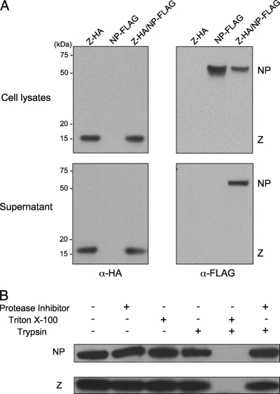
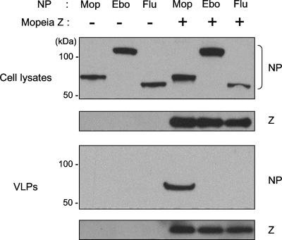
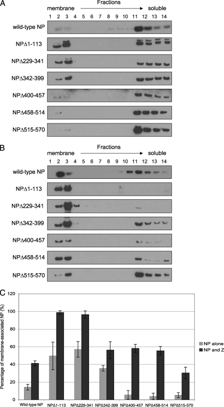
Arenaviruses are enveloped, negative-strand RNA viruses. For several arenaviruses, virus-like particle (VLP) formation requires the viral matrix Z protein. However, the mechanism by which viral ribonucleoprotein complexes are incorporated into virions is poorly understood. Here, we show that the expression of the Z protein and nucleoprotein (NP) of Mopeia virus, a close relative of the pathogenic Lassa virus, resulted in the highly selective incorporation of the NP protein into Z protein-induced VLPs. Moreover, the Z protein promoted the association of NP with cellular membranes, suggesting that the association of NP, Z, and the cellular membranes may facilitate the efficient incorporation of NP into VLPs. By employing a series of NP deletion constructs and testing their VLP incorporation, we further demonstrated an important role for the C-terminal half of NP in its incorporation into VLPs.


Figures

FIG. 1.
FIG. 1.
The Mopeia virus NP protein is incorporated into Z-induced VLPs. (A) pC-MopZ-HA, pC-MopNP-FLAG, or both plasmids were transfected into 293T cells. Forty-eight hours later, culture supernatants were cleared of debris, overlaid over a 20% sucrose cushion, and ultracentrifuged to pellet the VLPs. Cell lysates and corresponding pellets were analyzed by Western blotting with anti-HA and anti-FLAG antibodies. (B) Protease protection analysis of Mopeia virus VLPs. Vesicles isolated from the medium of pC-MopZ-HA- and pC-MopNP-FLAG-transfected 293T cells were divided into six aliquots. The six aliquots received the following treatments: (i) none, (ii) 3 mg/ml soybean trypsin inhibitor, (iii) 1% Triton X-100, (iv) 0.1 mg/ml trypsin, (v) 1% Triton X-100 and 0.1 mg/ml trypsin, and (vi) 3 mg/ml soybean trypsin inhibitor and 0.1 mg/ml trypsin. The samples were analyzed by Western blotting with anti-HA and anti-FLAG antibodies.
FIG. 2.
FIG. 2.
Selective incorporation of Mopeia virus NP into Z-driven VLPs. pC-MopNP-FLAG, pCEboZNPFLAG, or pC-WSN-FLAG-NP was transfected into 293T cells individually or in combination with pC-MopZ-HA. Forty-eight hours later, VLPs were isolated and analyzed by Western analysis with an anti-FLAG antibody. Mop, Mopeia virus NP; Ebo, Ebola virus NP; Flu, influenza A virus NP.
FIG. 3.
FIG. 3.
Mopeia virus NP colocalizes and redistributes with the Z protein. pC-MopNP-FLAG and pC-MopZ-HA were cotransfected into Vero cells. Twenty-four hours later, cells were fixed, permeabilized, and treated with anti-HA (green [FITC]) and anti-FLAG (red [Alexa Fluor 594]) antibodies. Confocal immunofluorescence microscopy was used to examine Z and NP fluorescence separately; the images were then merged to assess colocalization.
FIG. 4.
FIG. 4.
The Z protein promotes membrane association of the NP protein. 293T cells were transfected with pC-MopZ-HA/pC-eGFP, pC-MopNP-FLAG/pC-eGFP, or pC-MopNP-FLAG/pC-MopZ-HA/pC-eGFP. The cells were disrupted by passing them through a needle 48 h posttransfection. Cleared supernatants were then adjusted with the iodixanol solution OptiPrep to a final concentration of 40%, overlaid with 30% OptiPrep, and then overlaid with a buffer lacking OptiPrep and ultracentrifuged. The fractions were collected from the top of the tubes and analyzed by SDS-PAGE and Western blotting using either antibodies against alpha-calnexin and eGFP (A) or anti-HA and anti-FLAG antibodies to detect Mopeia virus Z and NP, respectively (B). eGFP served as a soluble, non-membrane-associated protein marker, and endogenous alpha-calnexin served as a membrane-associated protein marker.
FIG. 5.
FIG. 5.
Incorporation of NP mutants into VLPs. (A) Diagram of wild-type and NP deletion mutants. Constructs were fused with a FLAG tag at their C termini. The numbers indicate the deleted residues within wild-type NP. The results of protein expression and virion incorporation of mutant NPs are shown at the right. ND, not determined. (B) 293T cells were cotransfected with plasmids encoding wild-type or mutant NP and pC-MopZ-HA. After 48 h, VLPs were isolated and analyzed by Western blotting with anti-FLAG and anti-HA antibodies. The incorporation efficiency of mutant NP was calculated as the amount of NP divided by that of Z in the VLP sample. The incorporation efficiency for wild-type NP was set at 1, and the efficiencies for the mutant NPs were reported relative to the wild-type result. Three independent experiments were performed, and representative data are shown.
FIG. 6.
FIG. 6.
Effect of NP deletions on NP membrane association. The lysates of 293T cells expressing either an NP mutant and eGFP or an NP mutant, the Z protein, and eGFP were subjected to a membrane flotation assay, Western blot analysis and membrane association estimations are described in the legend to Fig. 4. (A and B) The results of Western blotting are shown for all mutants in the absence (A) or in the presence (B) of the Z protein. (C) The percentages of membrane-associated NP proteins were calculated as the ratio of the amount of NP in the calnexin-positive fractions to that in the whole gradient. The averages of data from three independent experiments are shown; the bars reflect the standard deviations.

Similar articles

Cited by

References

    1. Borden, K. L., E. J. Campbell Dwyer, and M. S. Salvato. 1998. An arenavirus RING (zinc-binding) protein binds the oncoprotein promyelocyte leukemia protein (PML) and relocates PML nuclear bodies to the cytoplasm. J. Virol. 72:758-766. - PMC - PubMed
    1. Briese, T., J. T. Paweska, L. K. McMullan, S. K. Hutchison, C. Street, G. Palacios, M. L. Khristova, J. Weyer, R. Swanepoel, M. Egholm, S. T. Nichol, and W. I. Lipkin. 2009. Genetic detection and characterization of Lujo virus, a new hemorrhagic fever-associated arenavirus from southern Africa. PLoS Pathog. 5:e1000455. - PMC - PubMed
    1. Buchmeier, M. J., J. C. de la Torre, and C. J. Peters. 2007. Arenaviridae, p. 1635-1668. In D. M. Knipe, P. M. Howley, D. E. Griffin, R. A. Lamb, M. A. Martin, B. Roizman, and S. E. Straus (ed.), Fields virology, 5th ed. Lippincott Williams & Wilkins, Philadelphia, PA.
    1. Burns, J. W., and M. J. Buchmeier. 1991. Protein-protein interactions in lymphocytic choriomeningitis virus. Virology 183:620-629. - PubMed
    1. Campbell Dwyer, E. J., H. Lai, R. C. MacDonald, M. S. Salvato, and K. L. Borden. 2000. The lymphocytic choriomeningitis virus RING protein Z associates with eukaryotic initiation factor 4E and selectively represses translation in a RING-dependent manner. J. Virol. 74:3293-3300. - PMC - PubMed

Publication types

LinkOut - more resources