. 2010 May;84(10):4969-78.
doi: 10.1128/JVI.02623-09. Epub 2010 Mar 3.

Binding site on the transferrin receptor for the parvovirus capsid and effects of altered affinity on cell uptake and infection

Affiliations

Binding site on the transferrin receptor for the parvovirus capsid and effects of altered affinity on cell uptake and infection

Laura B Goodman et al. J Virol. 2010 May.

Abstract

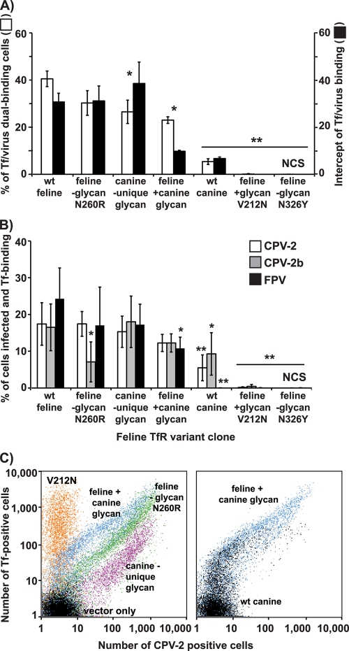
Canine parvovirus (CPV) and its relative feline panleukopenia virus (FPV) bind the transferrin receptor type 1 (TfR) to infect their host cells but show differences in the interactions with the feline and canine TfRs that determine viral host range and tissue tropism. We changed apical and protease-like domain residues by introducing point mutations and adding or removing glycosylation signals, and we then examined the interactions of those mutant TfRs with the capsids. Most substitutions had little effect on virus binding and uptake. However, mutations of several sites in the apical domain of the receptor either prevented binding to the capsids or reduced the affinity of receptor binding to various degrees. Glycans within the virus binding face of the apical domain also controlled capsid binding. CPV, but not the related feline parvovirus, could use receptors containing a canine TfR-specific glycosylation to mediate efficient infection, while addition of other N-linked glycosylation sites into the virus binding face of the feline apical domain reduced or eliminated both binding and infection. Replacement of critical feline TfR residue 221 with every amino acid had effects on binding and infection which were significantly associated with the biochemical properties of the residue replaced. Receptors with reduced affinities mostly showed proportional changes in their ability to mediate infection. Testing feline TfR variants for their binding and uptake patterns in cells showed that low-affinity versions bound fewer capsids and also differed in attachment to the cell surface and filopodia, but transport to the perinuclear endosome was similar.


Figures

FIG. 1.
FIG. 1.
(A) Locations of mutations tested, shown on a model of the feline sequence aligned to the human TfR structure. The different domains are colored in green (apical), red (protease-like), and yellow (helical). A portion of the stalk domain is colored in cyan (left), and the sites associated with Tf binding are brown (right). The following color code is used on the gray portion of the structure to highlight the substitutions which were made: red, Leu221; cyan, conserved N-linked glycosylation sites; orange, new glycosylation site added; pink, canine TfR unique glycosylation site added; dark blue, all other sites mutated in these studies. Changes with a large effect on virus binding are indicated by labels in red or purple. The labels of all the Asn-linked glycosylation sites are orange. (B) The position of residue Leu221 within the apical domain, shown in a model derived from the human TfR (the human position is Leu212). (C) Binding of CPV-2 capsids or infection by CPV-2 virus (as the percentage of positive cells) when using feline TfRs containing the indicated changes of residue 221, compared to the average buried accessible surface area (ASA) of the amino acid expressed. (D) Infection by CPV-2, CPV-2b, or FPV (shown as the percentage infection-positive cells) of cells expressing feline TfRs containing the indicated changes of residue 221, compared to average buried ASA of the amino acid expressed. (E) Binding of CPV-2 capsids to cells expressing feline TfRs containing the indicated changes of residue 221, compared to normalized van der Waals (VDW) side chain volumes of the replacement residues. Error bars indicate standard deviations.
FIG. 2.
FIG. 2.
Methods used for analyzing virus binding by the variant forms of the TfR. Representative examples of different binding profiles are shown, with Tf and virus (CPV-2) binding assayed by flow cytometry in transfected TRVb cells. The lines superimposed on the graphs represent regressions of the data in the upper right quadrant, and the intercept of the regression line with the axis between the Tf-positive and dual-stained quadrants shows the level of TfR expression (measured as Tf binding) required for detectable virus binding. Each panel represents three independent replicates of at least 10,000 cells, which were concatenated to make these plots.
FIG. 3.
FIG. 3.
Effects of replacements of various residues in the apical and protease-like domains of the feline TfR on the binding of virus and on virus infection, compared to those with the wild-type (wt) feline TfR. Means and standard deviations from three independent experiments are shown. Receptors not reaching the cell surface are indicated with the notation “NCS.” Statistically significant differences from the results seen for the wild-type feline TfR are noted (*, P < 0.05; **, P < 0.01); these were determined by testing the raw fluorescence data (not the mean values of percent bound or infected cells shown). (A) As illustrated in Fig. 2, flow cytometry was used to determine CPV-2 binding. Results of both analyses of viral binding are shown (percent dual CPV-2/Tf positive and intercept between the y axis of the Tf binding versus CPV-2 binding). The intercept represents the level of TfR expression on cells required to allow detectable virus attachment and uptake (here presented as the inverse for visual comparison) with cells not binding virus assigned an intercept of zero. (B) Infection of transfected TRVb cells with CPV-2, CPV-2b, and FPV. Cells were assayed by antibody staining combined with labeled Tf uptake and flow cytometry.
FIG. 4.
FIG. 4.
Effects of all replacements of residue 221 of the feline TfR on virus binding and virus infection. Virus binding (A) and infection (B) are shown as described in the legend of Fig. 3.
FIG. 5.
FIG. 5.
Characterization of feline and canine TfR mutants with alterations in glycosylation patterns, including either addition (V212N and K383N/S385T) or removal (N260R and N326Y) of sites within the apical domain, or the canine TfR with the unique glycosylation site (N383/T385) removed (30). Virus binding (A) and infection (B) are shown as described in the legend of Fig. 3, and representative binding profiles are compared (C).
FIG. 6.
FIG. 6.
Binding and uptake patterns of Tf or virus on TRVb cells expressing the wild-type feline TfR or the Ala221 mutant feline TfR, that shows a low affinity of virus attachment. (A) Cells after incubation with Tf for 15 or 30 min at 37°C, as indicated, and fixation with paraformaldehyde. A light microscopy image (phase) is shown, as well as a fluorescence image of labeled Tf (transferrin). (B) Cells after incubation with Alexa594-virus for 15 or 60 min at 37°C, as indicated, and fixation with paraformaldehyde. A light microscopy image (phase) is shown, as well as a fluorescence image of labeled capsids (virus). A magnified view shows an overlay of the cell phase image with the labeled capsids, with the association of virus with the filopodia on cells expressing the Ala221 TfR but not the wild type.

Similar articles

Cited by

References

    1. Abraham, J., K. D. Corbett, M. Farzan, H. Choe, and S. C. Harrison. 7 March 2010, posting date. Structural basis for receptor recognition by New World hemorrhagic fever arenaviruses. Nat. Struct. Mol. Biol. doi:10.1038/nsmb.1772. - DOI - PMC - PubMed
    1. Abraham, J., J. A. Kwong, C. G. Albarino, J. G. Lu, S. R. Radoshitzky, J. Salazar-Bravo, M. Farzan, C. F. Spiropoulou, and H. Choe. 2009. Host-species transferrin receptor 1 orthologs are cellular receptors for nonpathogenic New World clade B arenaviruses. PLoS Pathog. 5:e1000358. - PMC - PubMed
    1. Agbandje, M., R. McKenna, M. G. Rossmann, M. L. Strassheim, and C. R. Parrish. 1993. Structure determination of feline panleukopenia virus empty particles. Proteins 16:155-171. - PubMed
    1. Arnold, E., and M. G. Rossmann. 1990. Analysis of the structure of a common cold virus, human rhinovirus 14, refined at a resolution of 3.0 Å. J. Mol. Biol. 211:763-801. - PubMed
    1. Bates, G. W., and M. R. Schlabach. 1973. The reaction of ferric salts with transferrin. J. Biol. Chem. 248:3228-3232. - PubMed

Publication types

LinkOut - more resources