. 2010 May;84(10):4946-59.
doi: 10.1128/JVI.00060-10. Epub 2010 Mar 3.

The de novo methyltransferases DNMT3a and DNMT3b target the murine gammaherpesvirus immediate-early gene 50 promoter during establishment of latency

Affiliations

The de novo methyltransferases DNMT3a and DNMT3b target the murine gammaherpesvirus immediate-early gene 50 promoter during establishment of latency

Kathleen S Gray et al. J Virol. 2010 May.

Abstract

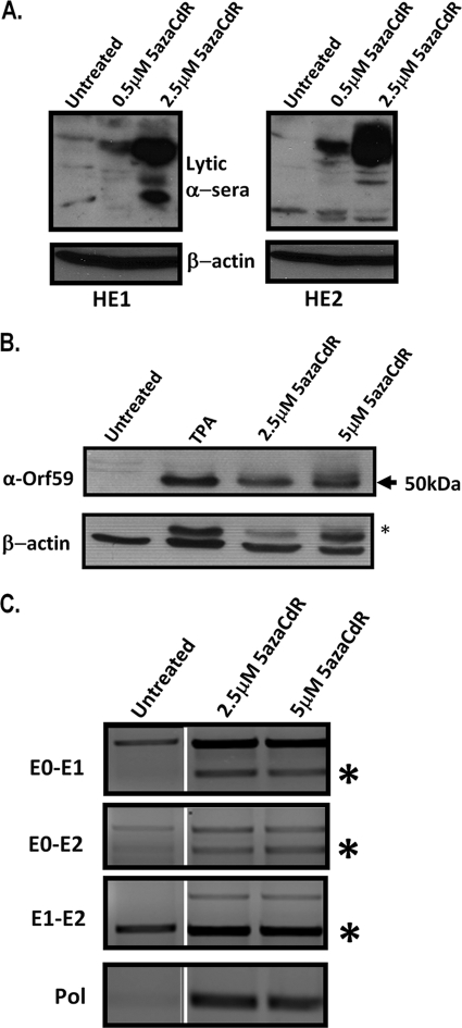
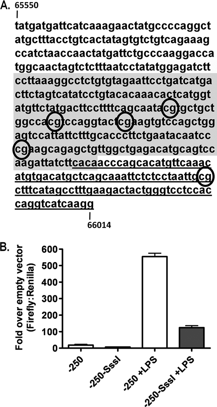
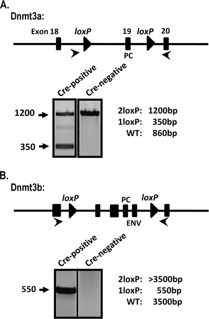
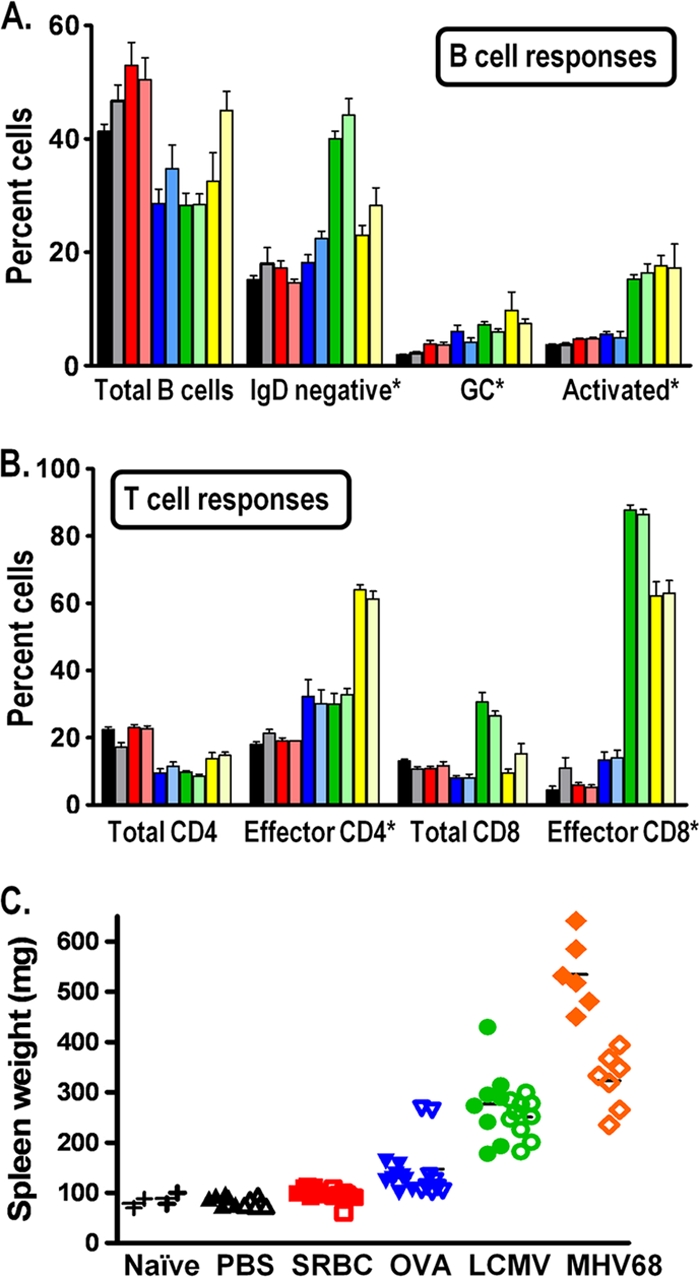
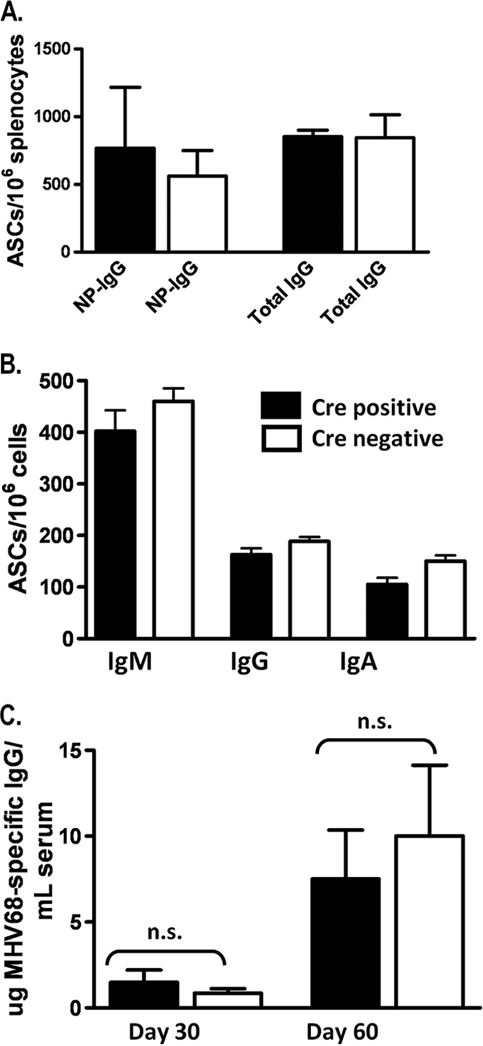
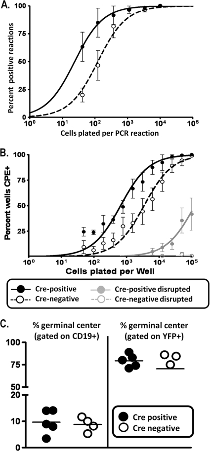
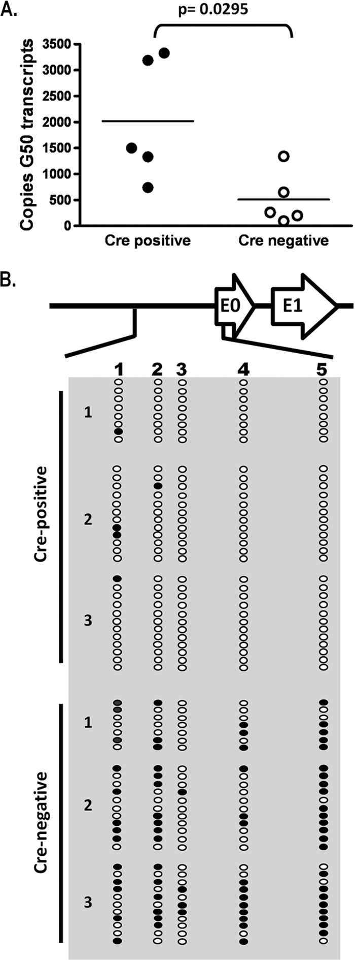
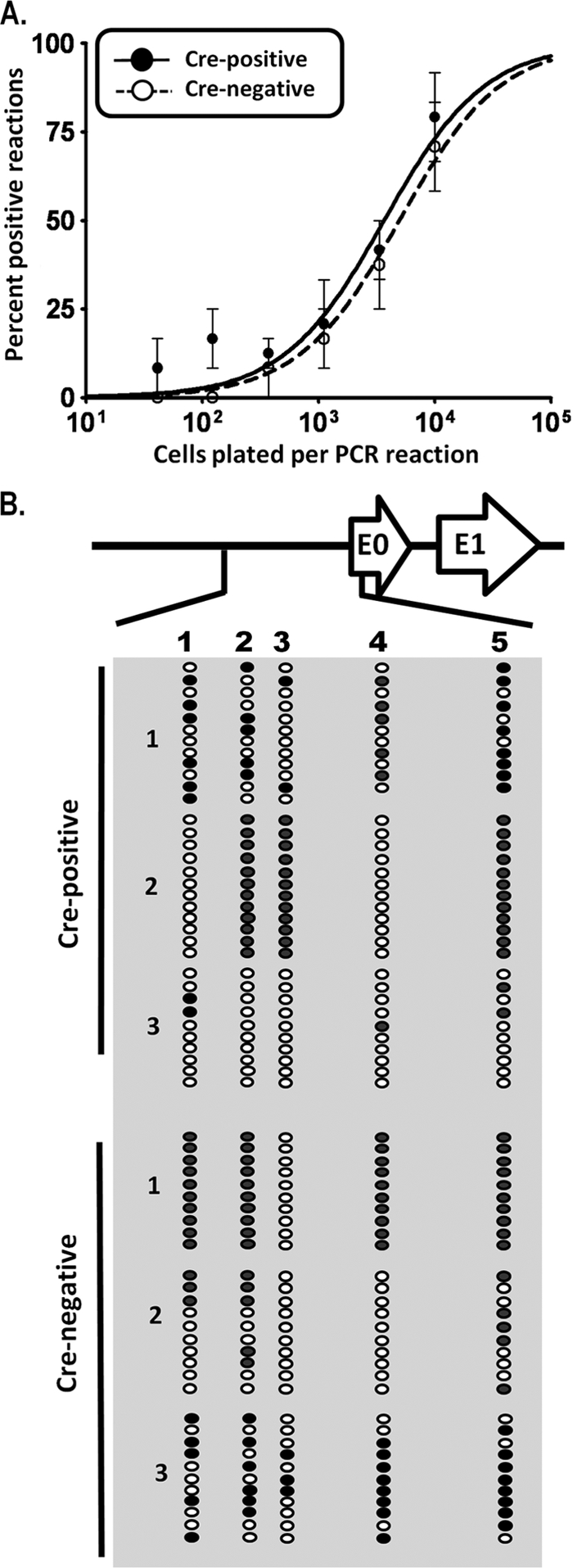
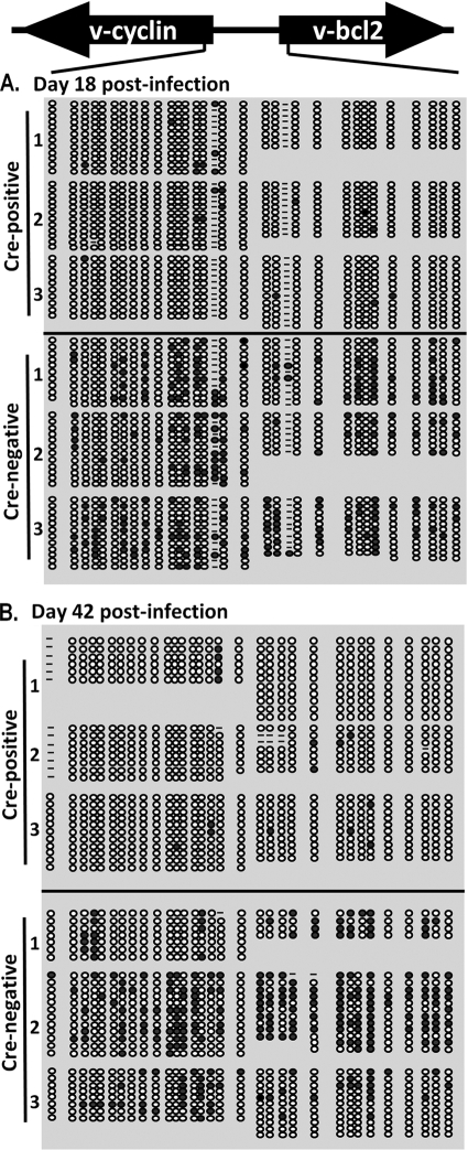
The role of epigenetic modifications in the regulation of gammaherpesvirus latency has been a subject of active study for more than 20 years. DNA methylation, associated with transcriptional silencing in mammalian genomes, has been shown to be an important mechanism in the transcriptional control of several key gammaherpesvirus genes. In particular, DNA methylation of the functionally conserved immediate-early replication and transcription activator (RTA) has been shown to regulate Epstein-Barr virus and Kaposi's sarcoma-associated herpesvirus Rta expression. Here we demonstrate that the murine gammaherpesvirus (MHV68) homolog, encoded by gene 50, is also subject to direct repression by DNA methylation, both in vitro and in vivo. We observed that the treatment of latently MHV68-infected B-cell lines with a methyltransferase inhibitor induced virus reactivation. In addition, we show that the methylation of the recently characterized distal gene 50 promoter represses activity in a murine macrophage cell line. To evaluate the role of de novo methyltransferases (DNMTs) in the establishment of these methylation marks, we infected mice in which conditional DNMT3a and DNMT3b alleles were selectively deleted in B lymphocytes. DNMT3a/DNMT3b-deficient B cells were phenotypically normal, displaying no obvious compromise in cell surface marker expression or antibody production either in naïve mice or in the context of nonviral and viral immunogens. However, mice lacking functional DNMT3a and DNMT3b in B cells exhibited hallmarks of deregulated MHV68 lytic replication, including increased splenomegaly and the presence of infectious virus in the spleen at day 18 following infection. In addition, total gene 50 transcript levels were elevated in the spleens of these mice at day 18, which correlated with the hypomethylation of the distal gene 50 promoter. However, by day 42 postinfection, aberrant virus replication was resolved, and we observed wild-type frequencies of viral genome-positive splenocytes in mice lacking functional DNMT3a and DNMT3b in B lymphocytes. The latter correlated with increased CpG methylation in the distal gene 50 promoter, which was restored to levels similar to those of littermate controls harboring functional DNMT3a and DNMT3b alleles in B lymphocytes, suggesting the existence of an alternative mechanism for the de novo methylation of the MHV68 genome. Importantly, this DNMT3a/DNMT3b-independent methylation appeared to be targeted specifically to the gene 50 promoter, as we observed that the promoters for MHV68 gene 72 (v-cyclin) and M11 (v-bcl2) remained hypomethylated at day 42 postinfection. Taken together, these data provide the first evidence of the importance of DNA methylation in regulating gammaherpesvirus RTA/gene 50 transcription during virus infection in vivo and provide insight into the hierarchy of host machinery required to establish this modification.


Figures

FIG. 1.
FIG. 1.
Methyltransferase inhibitor treatment induces MHV68 reactivation. (A) Whole-cell extracts from A20-HE1 or A20-HE2 cells treated for 72 h with 5azaCdR at the indicated concentrations and probed with rabbit-derived MHV68-lytic antisera. (B) Whole-cell extracts from A20-HE2 cells treated for 72 h with 5azaCdR at the indicated concentration and probed with Orf59 antibody. The asterisk in the beta-actin blot indicates residual Orf59 antibody that was not efficiently stripped prior to β-actin antibody hybridization. (C) A20-HE2 cells were treated for 72 h with the indicated concentrations of 5azaCdR. RT-PCR was performed to detect distal gene 50 promoter-initiated transcripts (containing E0) as well as E1 and E2 exon-containing transcripts. RT-PCR was also performed for a lytic transcript (viral DNA polymerase) as a verification of the induction of the MHV68 replication cycle. The asterisks indicate the band corresponding to the characterized spliced gene 50 transcripts (the larger amplification products detected represent either unspliced transcripts or contaminating viral DNA). The products from the RT-PCRs for both untreated and treated cells were electrophoresed in parallel on the same agarose gel alongside other samples unrelated to this experiment; these irrelevant lanes have been removed for presentation, and the amplification products arising from the untreated samples have been juxtaposed with those obtained with RNA prepared from 5azaCdR-treated cells.
FIG. 2.
FIG. 2.
In vitro methylation reduces distal gene 50 promoter activity. (A) Sequence of distal gene 50 promoter from bp 65550 to bp 66014. The CpGs examined by bisulfite analysis are indicated (numbered 1 to 5). Exon 0 is underlined, and the shaded region represents the −250 promoter region examined in B. (B) Reporter assay for distal gene 50 promoter activity in RAW264.7 murine macrophages with or without LPS (5 μg/ml) following M.SssI treatment or mock methylated. Data are representative of data from at least three independent transfections from two independent M.SssI treatments. P values were determined by using an unpaired Student's t test.
FIG. 3.
FIG. 3.
Rearrangement of Dnmt3a and Dnmt3b alleles in CD19+/Cre Dnmt3a/3bc/c splenocytes. Total genomic DNA was prepared from splenocytes from CD19+/Cre Dnmt3a/3bc/c (Cre-recombinase-positive) mice or littermate controls (Cre-recombinase negative). The schematic indicates the location of Dnmt3a or Dnmt3b exons (rectangles) and loxP sites (triangles). Cre-mediated rearrangement deletes exons encoding key DNMT catalytic domains: PC (Pro-Cys) and ENV (Glu-Asn-Val). PCR was performed with the primers indicated (arrows), and representative reactions are shown. WT, wild type.
FIG. 4.
FIG. 4.
Global immune responses are not altered in CD19+/Cre Dnmt3a/3bc/c mice. (A and B) B-cell (A) and T-cell (B) responses of CD19+/Cre Dnmt3a/3bc/c mice were examined. Splenocytes recovered from naïve mice (black/gray bars), mice immunized with SRBCs (day 18) (red bars) or OVA-CFA (day 14) (blue bars), or mice infected with LCMV Armstrong (day 8) (green bars) or MHV68 (day 18) (yellow bars) were examined by flow cytometry for the indicated populations. The darker bars correspond to cells recovered from mice lacking the expression of Dnmt3a and Dnmt3b in B cells (CD19+/Cre Dnmt3a/3bc/c mice), while the lighter bars indicate cells recovered from littermate control Dnmt3a/Dnmt3b-sufficient mice (CD19+/+ Dnmt3a/3bc/c mice). All populations were gated on live cells. The percentage of total B cells, total CD4+ T cells, and total CD8+ T cells are given relative to the numbers of total splenocytes. The asterisks in A and B indicate the gating of a specific population of splenocytes as follows: the percentages of specific B-cell populations (GC [germinal-center] B cells [GL7+/CD95+] and activated B cells [CD69high]) are given relative to numbers of total CD19+ cells (A), while the percentages of effector CD4+ T cells (CD44high/CD62Llow) and effector CD8+ T cells (CD44high/CD62Llow) are given relative to the numbers of either total CD4+ T cells or total CD8+ T cells, respectively (B). (C) Spleen weights from naïve mice or mice immunized or infected as in described above (A and B).
FIG. 5.
FIG. 5.
The antibody response of CD19+/Cre Dnmt3a/3bc/c mice is not grossly altered. (A) Numbers of total and NP-specific IgG-secreting B cells (from spleen) were determined by ELISPOT analysis 4 days following secondary immunization. (B) Numbers of IgM-, IgG-, or IgA-secreting cells were determined by ELISPOT analysis at day 18 following MHV68 infection. (C) MHV68-specific serum IgG levels were determined by ELISA at the indicated times postinfection. Data were analyzed by an unpaired Student's t test. n.s., not significant.
FIG. 6.
FIG. 6.
Evidence of ongoing lytic replication in CD19+/Cre Dnmt3a/3bc/c mice during early latency. (A) Limiting-dilution PCR analysis to determine frequencies of genome-positive cells. (B) Limiting-dilution analysis of intact and disrupted cells plated onto MEF monolayers to determine frequencies of cells reactivating from latency and to assess the contribution of preformed infectious virus to CPE. Genome and reactivation frequencies are elevated in Cre-positive mice, and the CPE observed upon the plating of disrupted cells indicates ongoing lytic replication. (C) Evidence that the MHV68-positive reservoir is not altered in CD19+/Cre Dnmt3a/3bc/c mice. Mice were infected with MHV68-YFP virus, and splenocytes were analyzed at day 18 for YFP expression and germinal-center markers (CD95 and GL7). The majority of YFP-positive (MHV68-infected) cells bear hallmarks of germinal-center B cells in both CD19+/Cre Dnmt3a/3bc/c and CD19+/+ Dnmt3a/3bc/c mice.
FIG. 7.
FIG. 7.
Elevated gene 50 transcription is accompanied by hypomethylation of the gene 50 promoter at day 18. (A) Quantitative RT-PCR for total gene 50 (G50) (exon 2) transcripts on cDNA generated from total splenocyte RNA. Transcript levels are elevated in Cre-positive mice relative to levels of Cre-negative littermate controls. P values were determined by using an unpaired Student's t test. (B) Bisulfite PCR analysis of the distal gene 50 promoter region at day 18. Total genomic DNA was extracted from magnetically purified CD19+ splenocytes from mice at day 18 postinfection and subjected to bisulfite PCR analysis. The region analyzed corresponds to bp 65550 to 66014 upstream of gene 50 exon 0 (Fig. 2). Each column represents an individual CpG dinucleotide, and each row represents an individual PCR clone. Filled circles represent methylated cytosines, while open circles represent unmethylated cytosines. Three individual mice were analyzed for Cre-positive and Cre-negative groups.
FIG. 8.
FIG. 8.
Normal genome frequency at day 42 postinfection is accompanied by restoration of gene 50 promoter methylation. (A) Limiting-dilution analysis to determine frequencies of genome-positive cells in Cre-positive and Cre-negative mice. Reactivation was negligible for both groups at this time. (B) Bisulfite PCR analysis of the distal gene 50 promoter region at day 42 postinfection. Total genomic DNA was extracted from purified CD19+ splenocytes isolated from mice at day 42 postinfection and subjected to bisulfite PCR analysis as described in the legend to Fig. 7.
FIG. 9.
FIG. 9.
Bisulfite PCR analysis of the v-cyclin and v-bcl2 promoter region at days 18 and 42 postinfection. Total genomic DNA was extracted from purified CD19+ splenocytes from mice at day 18 (A) or day 42 (B) postinfection and subjected to bisulfite PCR analysis. The region analyzed corresponds to bp 102548 to 103915, between the 5′ ends of each gene. Each column represents an individual CpG dinucleotide, and each row represents an individual PCR clone. Filled circles represent methylated cytosines, while open circles represent unmethylated cytosines. Dashes represent CpGs whose methylation status was indeterminate upon sequence analysis. Three individual mice were analyzed for Cre-positive and Cre-negative groups.

Similar articles

Cited by

References

    1. Allen, R. D., III, M. N. DeZalia, and S. H. Speck. 2007. Identification of an Rta responsive promoter involved in driving gammaHV68 v-cyclin expression during virus replication. Virology 365:250-259. - PMC - PubMed
    1. Allen, R. D., III, S. Dickerson, and S. H. Speck. 2006. Identification of spliced gammaherpesvirus 68 LANA and v-cyclin transcripts and analysis of their expression in vivo during latent infection. J. Virol. 80:2055-2062. - PMC - PubMed
    1. Barozzi, P., L. Potenza, G. Riva, D. Vallerini, C. Quadrelli, R. Bosco, F. Forghieri, G. Torelli, and M. Luppi. 2007. B cells and herpesviruses: a model of lymphoproliferation. Autoimmun. Rev. 7:132-136. - PubMed
    1. Ben-Sasson, S. A., and G. Klein. 1981. Activation of the Epstein-Barr virus genome by 5-aza-cytidine in latently infected human lymphoid lines. Int. J. Cancer 28:131-135. - PubMed
    1. Bhende, P. M., W. T. Seaman, H. J. Delecluse, and S. C. Kenney. 2005. BZLF1 activation of the methylated form of the BRLF1 immediate-early promoter is regulated by BZLF1 residue 186. J. Virol. 79:7338-7348. - PMC - PubMed

Publication types

LinkOut - more resources