. 2010 May;84(10):4979-87.
doi: 10.1128/JVI.01300-09. Epub 2010 Mar 3.

Regulation of Epstein-Barr virus origin of plasmid replication (OriP) by the S-phase checkpoint kinase Chk2

Affiliations

Regulation of Epstein-Barr virus origin of plasmid replication (OriP) by the S-phase checkpoint kinase Chk2

Jing Zhou et al. J Virol. 2010 May.

Abstract

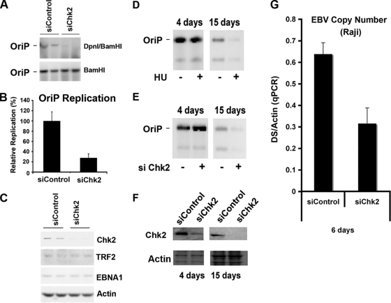
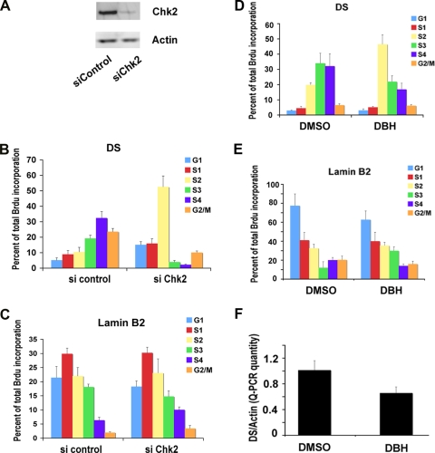
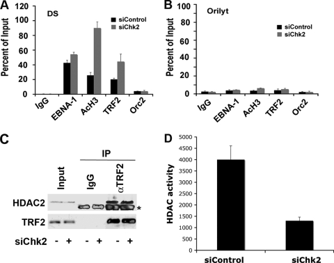
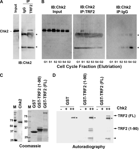
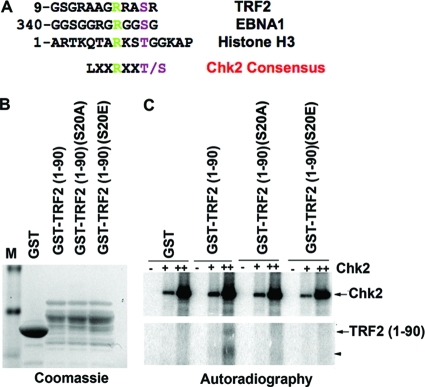
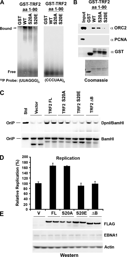
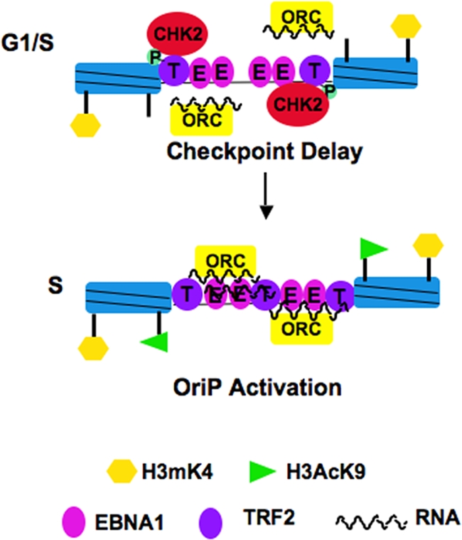
The Epstein-Barr virus (EBV) origin of plasmid replication (OriP) is required for episome stability during latent infection. Telomere repeat factor 2 (TRF2) binds directly to OriP and facilitates DNA replication and plasmid maintenance. Recent studies have found that TRF2 interacts with the DNA damage checkpoint protein Chk2. We show here that Chk2 plays an important role in regulating OriP plasmid stability, chromatin modifications, and replication timing. The depletion of Chk2 by small interfering RNA (siRNA) leads to a reduction in DNA replication efficiency and a loss of OriP-dependent plasmid maintenance. This corresponds to a change in OriP replication timing and an increase in constitutive histone H3 acetylation. We show that Chk2 interacts with TRF2 in the early G(1)/S phase of the cell cycle. We also show that Chk2 can phosphorylate TRF2 in vitro at a consensus acceptor site in the amino-terminal basic domain of TRF2. TRF2 mutants with a serine-to-aspartic acid phosphomimetic substitution mutation were reduced in their ability to recruit the origin recognition complex (ORC) and stimulate OriP replication. We suggest that the Chk2 phosphorylation of TRF2 is important for coordinating ORC binding with chromatin remodeling during the early S phase and that a failure to execute these events leads to replication defects and plasmid instability.


Figures

FIG. 1.
FIG. 1.
Chk2 depletion inhibits OriP DNA replication and plasmid maintenance. (A) HCT116 cells were cotransfected with siControl or siChk2 siRNA and with OriP plasmid-coexpressing EBNA1 and assayed for transient DNA replication. Plasmid DNA, shown in duplicates, was digested with BamHI (bottom) or DpnI plus BamHI (bottom) and visualized by Southern blotting. (B) Replication efficiency was quantified for at least three independent transfections as the ratio of resistant DpnI to total plasmid DNA recovered (BamHI-linearized DNA), and replication for siCon was normalized to 100%. (C) Western blot control to assay siChk2 depletion and EBNA1, TRF2, and actin expression levels in cells shown in A. (D) HeLa cells were selected for GFP-expressing OriP plasmids and then treated with (+) or without (−) HU (50 μM) for 4 or 15 days, as indicated. OriP plasmids were then extracted from the same number of cells and assayed by Southern blotting. (E) Same as above (D) except that cells were transfected with control or Chk2-targeting siRNA every 3 days for 4 or 15 days. No HU was included in these experiments. (F) The siRNA-transfected cells shown in E were analyzed by Western blotting for Chk2 (top) or actin (bottom) at 4 and 15 days after transfection of the OriP plasmid, as indicated. (G) Raji cells were nucleofected with siRNA for Chk2 or control siRNA every third day for 6 days. The EBV genome copy number was measured by real-time PCR analysis of the EBV DS region relative to cellular actin DNA. qPCR, quantitative PCR.
FIG. 2.
FIG. 2.
Chk2 depletion or Chk2 inhibition by DBH alters OriP replication timing and function. (A) D98/HR1 cells were transfected with siControl or siChk2 RNA and assayed by Western blotting to verify that Chk2 was depleted at 72 h posttransfection. (B and C) siControl- or siChk2-transfected D98/HR1 cells were pulse-labeled with BrdU, stained with PI, and then fractionated into different cell cycle stages by using FACS. BrdU-labeled DNA specific for the DS (B) or cellular lamin B2 (C) was quantified by anti-BrdU immunoprecipitation and PCR. Cell cycle fractions are indicated. (D and E) A replication timing assay was performed with D98/HR1 cells treated with DBH or control DMSO for 48 h prior to BrdU pulse-labeling, FACS, and IP-PCR analysis. BrdU incorporation was assayed at the EBV DS (D) or at the cellular lamin B2 origin (E). (F) EBV genome copy numbers were assayed in D98/HR1 cells treated with DBH or the DMSO control after 6 days of treatment. The copy number was measured by real-time PCR analysis of the EBV DS relative to cellular actin DNA.
FIG. 3.
FIG. 3.
Chk2 depletion abrogates histone deacetylation at the DS. (A and B) D98/HR1 cells were transfected with siControl or siChk2 RNA and then assayed by ChIP for binding of EBNA1, AcH3, TRF2, ORC2, or control IgG at EBV regions for the DS (A) or OriLyt (B). ChIP DNA was quantified by real-time PCR as a relative percentage of the input. (C) D98/HR1 cells were transfected with siChk2 (+) or siControl (−) and then assayed for HDAC2 abundance by Western blotting (input) or after immunoprecipitation with IgG control or anti-TRF2 antibody, as indicated. Control Western blots for TRF2 are indicated at the bottom. The asterisk indicates a cross-reacting IgG band from the immunoprecipitating antibody. (D) HDAC activity assay from total cell extracts derived from siControl- or siChk2-transfected D98/HR1 cells.
FIG. 4.
FIG. 4.
Chk2 binds TRF2 in a cell cycle-dependent manner and phosphorylates TRF2 in vitro. (A) Asynchronous Raji cell cultures were subjected to immunoprecipitation with antibodies to TRF2 or control IgG and then assayed by Western blotting for Chk2. Input lysates (shown in the left lane) represent 10% of starting material for IP. IB, immunoblot. (B) Raji cells were cell cycle fractionated using centrifugal elutriation and then subjected to IP with antibodies to TRF2 or control IgG, as indicated. Input lanes represent 10% of starting material. The amount of the Chk2 protein was assayed by Western blotting with anti-Chk2 antibody. The asterisks indicate the nonspecific cross-reaction with the IgG heavy and light chains from IP. (C) Coomassie blue staining of SDS-PAGE gels was used to assess the purity of the baculovirus-derived Chk2 kinase (left) or the bacterially derived GST, GST-TRF2(1-90), and GST-TRF2(FL) proteins used for kinase assays. (D) The purified Chk2 protein was assayed at 0 nM, 50 nM (+), or 200 nM (++) for its ability to phosphorylate GST, GST-TRF2(1-90), or GST-TRF2(FL), as indicated. 32P-labeled TRF2 fusion proteins are indicated by arrows, and the arrowhead indicates a GST-TRF2 breakdown product. Chk2 autophosphorylation is indicated by asterisks.
FIG. 5.
FIG. 5.
Serine 20 in TRF2 can be phosphorylated by Chk2 in vitro. (A) Candidate Chk2 phosphorylation sites in TRF2 (S20), EBNA1 (S350), and histone H3 (T11) are aligned with the known consensus sites for Chk2 phosphorylation. (B) GST, GST-TRF2(1-90), GST-TRF2(1-90)(S20A), and GST-TRF2(1-90)(S20E) were purified from E. coli cells and visualized by Coomassie blue staining after SDS-PAGE. (C) The purified Chk2 protein at 0 nM, 50 nM (+), or 250 nM (++) was assayed for its ability to phosphorylate GST, GST-TRF2(1-90), GST-TRF2(1-90)(S20A), and GST-TRF2(1-90)(S20E), as indicated. The arrows indicate TRF2(1-90), and the arrowhead indicates a GST-TRF2 breakdown product. Chk2 autophosphorylation is indicated in the upper panel.
FIG. 6.
FIG. 6.
TRF2 phosphomimetic mutant S20E abrogates RNA binding, ORC recruitment, and OriP DNA replication. (A) GST, GST-TRF2(1-90), GST-TRF2(1-90)(S20A), and GST-TRF2(1-90)(S20E) were assayed by EMSA for RNA binding to a G-rich probe, (UUAGGG)8 (left), or a C-rich probe, (CCCUAA)8 (right). (B) GST, GST-TRF2(1-90), GST-TRF2(1-90)(S20A), and GST-TRF2(1-90)(S20E) were incubated with HeLa nuclear extracts (0.5 ml at 5 mg/ml), washed extensively, and then assayed by SDS-PAGE for the recruitment of ORC2 (top) or control PCNA (middle). Loading controls for GST (bottom) or Coomassie staining is indicated below. Std, standard. (C) Transient DNA replication assays were performed with HCT116 cells with an OriP plasmid coexpressing the EBNA1 protein. Cells were cotransfected with a cytomegalovirus (CMV)-Flag vector or CMV-FLAG-TRF2(FL) or the S20A, S20E, or ΔB mutant and then assayed at 72 h posttransfection for OriP replication. Recovered plasmid DNA was digested with DpnI plus BamHI (top) or BamHI alone (bottom) and assayed by Southern blotting. (D) Quantification of at least three independent transfection assays represented in C. (E) Western blot analysis of FLAG-TRF2 proteins (anti-FLAG) transfected for the replication assays shown in C. EBNA1 and actin served as transfection and loading controls, respectively.
FIG. 7.
FIG. 7.
Speculative model for Chk2 regulation of OriP. Chk2 is shown to associate with phosphorylated TRF2 in the G1/S phase. Phosphorylated TRF2 is compromised in RNA binding and ORC recruitment, which may prevent replication initiation and provide a Chk2-dependent checkpoint delay in replication initiation. In the mid-S phase, Chk2 dissociates from TRF2 and returns OriP to a competent replication origin.

Similar articles

Cited by

References

    1. Atanasiu, C., Z. Deng, A. Wiedmer, J. Norseen, and P. M. Lieberman. 2006. ORC binding to TRF2 stimulates OriP replication. EMBO Rep. 7:716-721. - PMC - PubMed
    1. Bashaw, J. M., and J. L. Yates. 2001. Replication from oriP of Epstein-Barr virus requires exact spacing of two bound dimers of EBNA1 which bend DNA. J. Virol. 75:10603-10611. - PMC - PubMed
    1. Bell, S. P. 2002. The origin recognition complex: from simple origins to complex functions. Genes Dev. 16:659-672. - PubMed
    1. Bhaskara, S., B. J. Chyla, J. M. Amann, S. K. Knutson, D. Cortez, Z. W. Sun, and S. W. Hiebert. 2008. Deletion of histone deacetylase 3 reveals critical roles in S phase progression and DNA damage control. Mol. Cell 30:61-72. - PMC - PubMed
    1. Buscemi, G., L. Zannini, E. Fontanella, D. Lecis, S. Lisanti, and D. Delia. 2009. The shelterin protein TRF2 inhibits Chk2 activity at telomeres in the absence of DNA damage. Curr. Biol. 19:874-879. - PubMed

Publication types