

Activation of tolerogenic dendritic cells in the tumor draining lymph nodes by CD8+ T cells engineered to express CD40 ligand
- PMID: 20200275
- PMCID: PMC2843821
- DOI: 10.4049/jimmunol.0903111
Activation of tolerogenic dendritic cells in the tumor draining lymph nodes by CD8+ T cells engineered to express CD40 ligand
Abstract
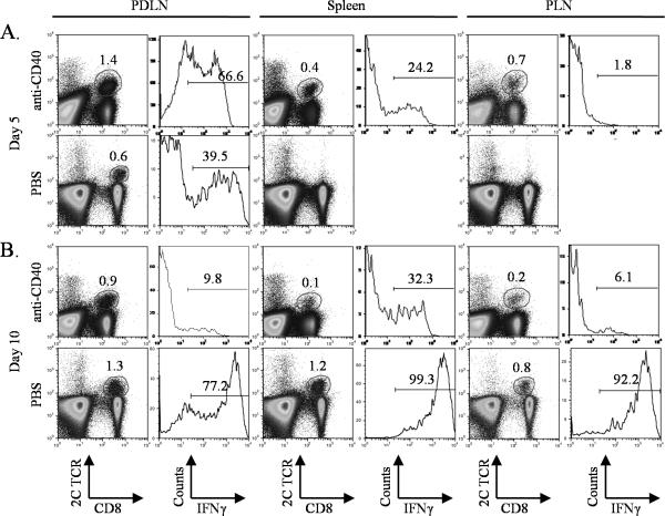
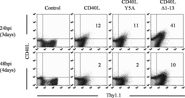
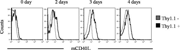
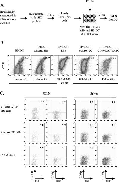
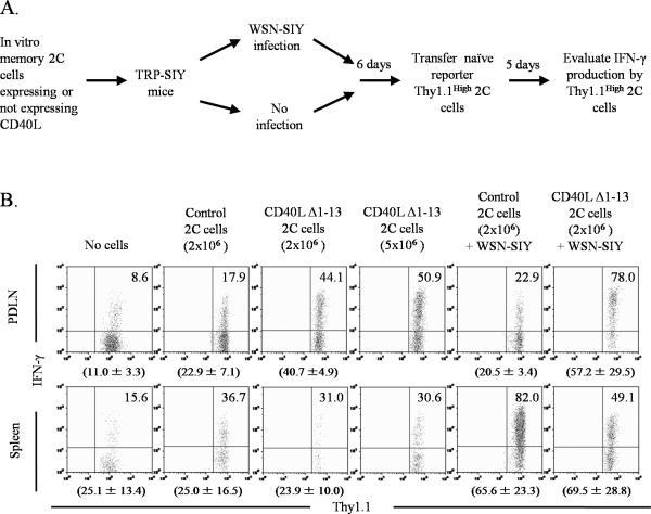
Tolerogenic dendritic cells in the tumor microenvironment can inhibit the generation and maintenance of robust antitumor T cell responses. In this study, we investigated the effects of local delivery of CD40L by tumor-reactive CD8(+) T cells on dendritic cell activation and antitumor T cell responses in the TRAMP model. To increase the immunostimulatory signal, CD40L was engineered, by deleting the majority of the cytoplasmic domain, to increase its levels of expression and duration on the surface of CD8(+) T cells. Tumor-reactive CD8(+) T cells expressing the truncated form of CD40L stimulated maturation of dendritic cells in vitro and in the prostate draining lymph nodes in vivo. Following dendritic cell maturation, a significantly higher fraction of adoptively transferred, tumor-reactive (reporter) CD8(+) T cells was stimulated to express IFN-gamma and infiltrate the prostate tissue. The antitumor CD8(+) T cell response was further enhanced if TRAMP mice were also immunized with a tumor-specific Ag. These findings demonstrate that augmented T cell responses can be achieved by engineering tumor-reactive T cells to deliver stimulatory signals to dendritic cells in the tumor microenvironment.
Figures
Similar articles
-
CD40 ligand promotes priming of fully potent antitumor CD4(+) T cells in draining lymph nodes in the presence of apoptotic tumor cells.J Immunol. 2001 Nov 15;167(10):5678-88. doi: 10.4049/jimmunol.167.10.5678. J Immunol. 2001. PMID: 11698440
-
Direct T cell activation via CD40 ligand generates high avidity CD8+ T cells capable of breaking immunological tolerance for the control of tumors.PLoS One. 2014 Mar 24;9(3):e93162. doi: 10.1371/journal.pone.0093162. eCollection 2014. PLoS One. 2014. PMID: 24664420 Free PMC article.
-
Reinforcement of cancer immunotherapy by adoptive transfer of cblb-deficient CD8+ T cells combined with a DC vaccine.Immunol Cell Biol. 2012 Jan;90(1):130-4. doi: 10.1038/icb.2011.11. Epub 2011 Mar 8. Immunol Cell Biol. 2012. PMID: 21383769
-
Immunity to murine prostatic tumors: continuous provision of T-cell help prevents CD8 T-cell tolerance and activates tumor-infiltrating dendritic cells.Cancer Res. 2009 Aug 1;69(15):6256-64. doi: 10.1158/0008-5472.CAN-08-4516. Epub 2009 Jul 21. Cancer Res. 2009. Retraction in: Cancer Res. 2016 Apr 15;76(8):2490. doi: 10.1158/0008-5472.CAN-16-0505. PMID: 19622771 Free PMC article. Retracted.
-
Dendritic cell gene therapy.Surg Oncol Clin N Am. 2002 Jul;11(3):645-60. doi: 10.1016/s1055-3207(02)00027-3. Surg Oncol Clin N Am. 2002. PMID: 12487060 Review.
Cited by
-
Dendritic cell-based vaccine efficacy: aiming for hot spots.Front Immunol. 2015 Mar 3;6:91. doi: 10.3389/fimmu.2015.00091. eCollection 2015. Front Immunol. 2015. PMID: 25784913 Free PMC article. Review.
-
Immune-based antitumor effects of BRAF inhibitors rely on signaling by CD40L and IFNγ.Cancer Res. 2014 Jun 15;74(12):3205-17. doi: 10.1158/0008-5472.CAN-13-3461. Epub 2014 Apr 15. Cancer Res. 2014. PMID: 24736544 Free PMC article.
-
Double Strike Approach for Tumor Attack: Engineering T Cells Using a CD40L:CD28 Chimeric Co-Stimulatory Switch Protein for Enhanced Tumor Targeting in Adoptive Cell Therapy.Front Immunol. 2021 Nov 29;12:750478. doi: 10.3389/fimmu.2021.750478. eCollection 2021. Front Immunol. 2021. PMID: 34912334 Free PMC article.
-
T cells as vehicles for cancer vaccination.J Biomed Biotechnol. 2011;2011:417403. doi: 10.1155/2011/417403. Epub 2011 Oct 27. J Biomed Biotechnol. 2011. PMID: 22131805 Free PMC article. Review.
-
Enhancing antitumor efficacy of chimeric antigen receptor T cells through constitutive CD40L expression.Mol Ther. 2015 Apr;23(4):769-78. doi: 10.1038/mt.2015.4. Epub 2015 Jan 13. Mol Ther. 2015. PMID: 25582824 Free PMC article.
References
-
- Dudley ME, Wunderlich JR, Robbins PF, Yang JC, Hwu P, Schwartzentruber DJ, Topalian SL, Sherry R, Restifo NP, Hubicki AM, Robinson MR, Raffeld M, Duray P, Seipp CA, Rogers-Freezer L, Morton KE, Mavroukakis SA, White DE, Rosenberg SA. Cancer regression and autoimmunity in patients after clonal repopulation with antitumor lymphocytes. Science. 2002;298:850–854. - PMC - PubMed
-
- Dudley ME, Wunderlich JR, Yang JC, Sherry RM, Topalian SL, Restifo NP, Royal RE, Kammula U, White DE, Mavroukakis SA, Rogers LJ, Gracia GJ, Jones SA, Mangiameli DP, Pelletier MM, Gea-Banacloche J, Robinson MR, Berman DM, Filie AC, Abati A, Rosenberg SA. Adoptive cell transfer therapy following non-myeloablative but lymphodepleting chemotherapy for the treatment of patients with refractory metastatic melanoma. J. Clin. Oncol. 2005;23:2346–2357. - PMC - PubMed
-
- Dudley ME, Yang JC, Sherry R, Hughes MS, Royal R, Kammula U, Robbins PF, Huang J, Citrin DE, Leitman SF, Wunderlich J, Restifo NP, Thomasian A, Downey SG, Smith FO, Klapper J, Morton K, Laurencot C, White DE, Rosenberg SA. Adoptive cell therapy for patients with metastatic melanoma: evaluation of intensive myeloablative chemoradiation preparative regimens. J. Clin. Oncol. 2008;26:5233–5239. - PMC - PubMed
-
- Zou W. Immunosuppressive networks in the tumour environment and their therapeutic relevance. Nat. Rev. Cancer. 2005;5:263–274. - PubMed
-
- Chomarat P, Banchereau J, Davoust J, Palucka AK. IL-6 switches the differentiation of monocytes from dendritic cells to macrophages. Nat. Immunol. 2000;1:510–514. - PubMed
Publication types
MeSH terms
Substances
Grants and funding
LinkOut - more resources
Full Text Sources
Other Literature Sources
Research Materials
