. 2010 Jun;59(6):1312-20.
doi: 10.2337/db09-1155. Epub 2010 Mar 3.

PTEN inhibition improves muscle regeneration in mice fed a high-fat diet

Affiliations

PTEN inhibition improves muscle regeneration in mice fed a high-fat diet

Zhaoyong Hu et al. Diabetes. 2010 Jun.

Abstract

Objective: Mechanisms impairing wound healing in diabetes are poorly understood. To identify mechanisms, we induced insulin resistance by chronically feeding mice a high-fat diet (HFD). We also examined the regulation of phosphatidylinositol 3,4,5-trisphosphate (PIP(3)) during muscle regeneration because augmented IGF-1 signaling can improve muscle regeneration.

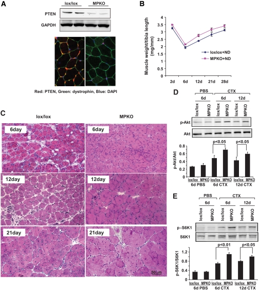
Research design and methods: Muscle regeneration was induced by cardiotoxin injury, and we evaluated satellite cell activation and muscle maturation in HFD-fed mice. We also measured PIP(3) and the enzymes regulating its level, IRS-1-associated phosphatidylinositol 3-kinase (PI3K) and PTEN. Using primary cultures of muscle, we examined how fatty acids affect PTEN expression and how PTEN knockout influences muscle growth. Mice with muscle-specific PTEN knockout were used to examine how the HFD changes muscle regeneration.

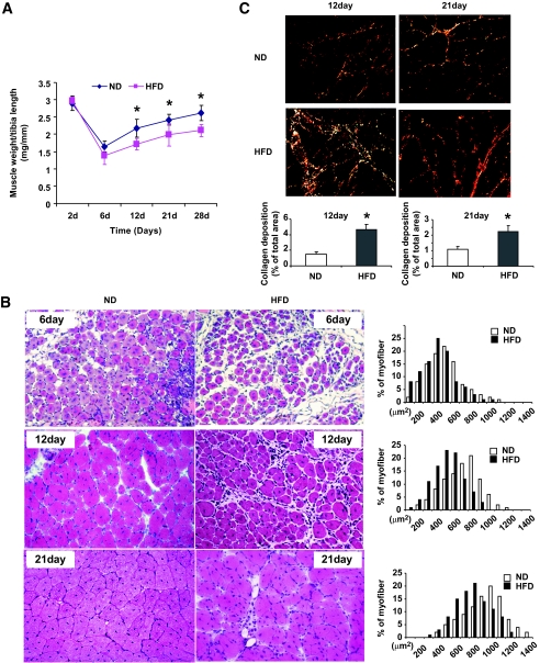
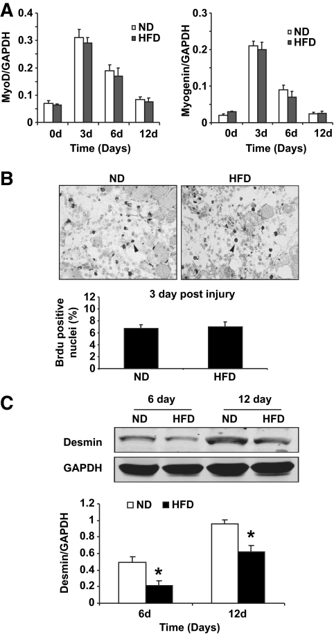
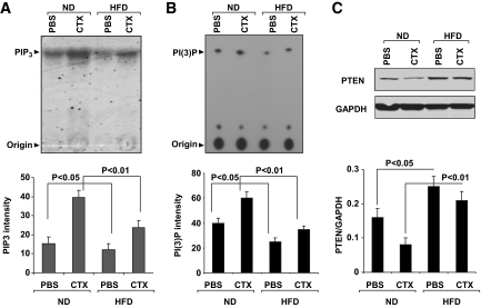
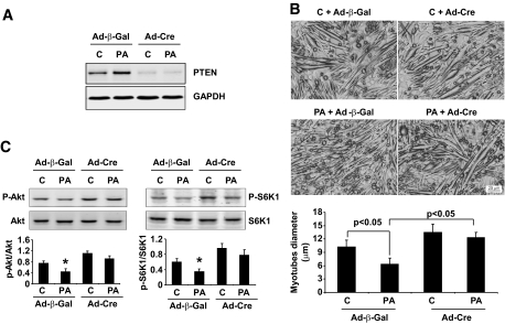
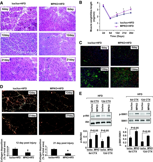
Results: The HFD raised circulating fatty acids and impaired the growth of regenerating myofibers while delaying myofiber maturation and increasing collagen deposition. These changes were independent of impaired proliferation of muscle progenitor or satellite cells but were principally related to increased expression of PTEN, which reduced PIP(3) in muscle. In cultured muscle cells, palmitate directly stimulated PTEN expression and reduced cell growth. Knocking out PTEN restored cell growth. In mice, muscle-specific PTEN knockout improved the defects in muscle repair induced by HFD.

Conclusions: Insulin resistance impairs muscle regeneration by preventing myofiber maturation. The mechanism involves fatty acid-stimulated PTEN expression, which lowers muscle PIP(3). If similar pathways occur in diabetic patients, therapeutic strategies directed at improving the repair of damaged muscle could include suppression of PTEN activity.


Figures

FIG. 1.
FIG. 1.
An HFD delays the recovery of and increases fibrosis in regenerating muscle. A: The weights of injured tibialis anterior muscles (normalized to the length of the tibia) were decreased in mice fed the HFD for 8 months. Muscles were obtained at 6, 12, 21, and 28 days after injury and compared with results from mice fed the normal diet (ND). n = 12 in each group. B: Hematoxylin-eosin sections of injured tibialis anterior muscles from HFD (right panel) and normal diet (left panel) mice revealed smaller regenerating myofibers (detected by their central nuclei). There also was an increase in interstitial space of muscle. The distribution of cross-sectional areas of new myofibers was shifted leftwards compared with values in mice fed a normal diet. C: Sirius red staining revealed an increase in collagen deposition at 12 (left panel) and 21 (right panel) days after injury in HFD mice. The collagen-containing area was significantly increased in muscles of HFD mice compared with results from muscles of mice on a normal diet (n = 6 in each group). *P < 0.01. d, days. (A high-quality digital representation of this figure is available in the online issue.)
FIG. 2.
FIG. 2.
An HFD suppresses maturation of myofibers in regenerating muscle. A: The mRNAs of MyoD or myogenin (markers of satellite cell proliferation) in injured muscles of mice fed the HFD were no different from those in mice fed the normal diet (ND) at days 3 and 6 after injury (n = 6 in each group). B: Cell proliferation at day 3 after injury was assessed by measuring Brdu incorporation. Arrows indicated typical Brdu-positive nuclei. There were no statistical differences in values from HFD (right panel) and normal diet (left panel) mice (n = 6 in each group). d, days. C: At day 6 and 12 after injury, Western blotting revealed significantly decreased desmin expression in muscles of HFD mice (n = 6 in each group of mice) *P < 0.05). d, days.
FIG. 3.
FIG. 3.
HFD suppresses the accumulation of PIP3 and IRS-1–associated PI3K activity in muscle during regeneration. A: At day 6 after injury, PIP3 was increased in regenerating muscles of mice fed a normal diet (ND); the HFD reduced PIP3 in uninjured and injured muscles (n = 6 in each group). B: In muscles of ND mice, IRS-1–associated PI3K activity was increased at day 6 after injury; the HFD significantly decreased this activity in uninjured and injured muscles (n = 6 in each group). C: In mice mice on a normal diet at 6 days after injury, Western blotting revealed a decrease in PTEN in regenerating myofibers. The HFD stimulated PTEN expression compared with results in mice on a normal diet (n = 6 in each group). CTX, cardiotoxin injury.
FIG. 4.
FIG. 4.
Palmitate impairs the growth of cultured myotubes, and PTEN deletion prevents it. A: Primary cultures of muscle cells from PTENlox/lox Cre mice were infected by adenoviruses: Ad-Cre to delete PTEN or Ad-β-Gal (control). Infected cells were then treated with palmitate (PA) or vehicle (C), and the expression of PTEN was evaluated by Western blot using GAPDH as the loading control. B: Cultured muscle cells from A were examined for difference in myotube diameters. C: Phosphorylation of Akt and S6K1 was evaluated in muscle cell cultures from A. The ratios of p-Akt/Akt (Ser473) and p-S6K1/S6K1 (Thr389) were calculated for three separate experiments.
FIG. 5.
FIG. 5.
Muscle-specific PTEN deletion (MPKO) improved muscle regeneration in 3-month-old mice fed a normal diet. A: PTEN protein was markedly decreased in MPKO muscles vs. control lox/lox mice as assessed by Western blot (upper panel). PTEN (red) immunostaining (lower panel) was absent in myofibers of MPKO mice but was present in vascular or satellite cells (arrow). Green, dystrophin. Blue, DAPI. B: The weight of tibialis anterior muscles (factored by tibia length) after injury was improved in MPKO mice fed a normal diet compared with that in HFD mice (n = 6 in each group). C: Hematoxylin-eosin staining of tibialis anterior injured muscles in MPKO mice fed a normal diet revealed larger myofibers with central nuclei compared with responses in lox/lox mice. D: At 6 and 12 days after cardiotoxin (CTX) injury, the ratio of p-Akt/Akt (Ser473) was significantly higher in muscle of MPKO vs. lox/lox mice. Both groups were fed the normal diet (P < 0.05; n = 6 in each group). E: the increase in p-Akt was associated with an increase in p-S6K1 (Thr389). Total S6K1 was used as loading control (P < 0.05; n = 6 in each group). *P < 0.05. d, days. (A high-quality digital representation of this figure is available in the online issue.)
FIG. 6.
FIG. 6.
In HFD mice with muscle-specific PTEN deletion (MPKO), the suppression of myofiber maturation and the increase in collagen deposition were blocked. A: Hematoxylin-eosin staining revealed improved maturation of myofibers in injured tibialis anterior muscles of MPKO HFD mice (right panel) compared with results in muscles of HFD control lox/lox mice (left panel). B: MPKO improved weight gain of injured tibialis anterior muscles factored for tibia length compared with results from lox/lox HFD mice (n = 12 in each group). *P < 0.01. C: At 6 and 12 days after injury, immunostaining of the differentiation marker, desmin, increased in injured muscles of MPKO mice fed the HFD (right panel) at 6 and 12 days after injury compared with results in HFD control lox/lox mice (left panel). D: At days 12 and 21 after injury, Sirius red staining for collagen in muscles of HFD MPKO mice (right panel) revealed a significant reduction vs. results in muscles of HFD lox/lox mice (left panel). Bar graphs (lower panel) represent the fractions of injured muscle staining for collagen. n = 6. *P < 0.05. E: The ratio of p-Akt/Akt (Ser473) in muscle of lox/lox mice fed the HFD was lower than in muscle of MPKO mice (n = 6 in each group). The ratio of p-S6K1/S6K1 (Thr389) had a similar pattern (n = 6 in each group). CTX, cardiotoxin injury. (A high-quality digital representation of this figure is available in the online issue.)

Similar articles

Cited by

References

    1. Hirsch T, Spielmann M, Velander P, Zuhaili B, Bleiziffer O, Fossum M, Steinstraesser L, Yao F, Eriksson E: Insulin-like growth factor-1 gene therapy and cell transplantation in diabetic wounds. J Gene Med 2008; 10: 1247–1252 - PubMed
    1. Vignaud A, Ramond F, Hourde C, Keller A, Butler-Browne G, Ferry A: Diabetes provides an unfavorable environment for muscle mass and function after muscle injury in mice. Pathobiology 2007; 74: 291–300 - PubMed
    1. Shortreed K, Johnston A, Hawke TJ: Satellite cells and muscle repair. In Skeletal Muscle Damage and Repair 1st ed Tiidus PM: Ed. Champaign, IL, Human Kinetics, 2008, p. 77–88
    1. Grefte S, Kuijpers-Jagtman AM, Torensma R, Von den Hoff JW: Skeletal muscle development and regeneration. Stem Cells Dev 2007; 16: 857–868 - PubMed
    1. Charge SB, Rudnicki MA: Cellular and molecular regulation of muscle regeneration. Physiol Rev 2004; 84: 209–238 - PubMed

Publication types

MeSH terms