

Implication of ethanol wet-bonding in hybrid layer remineralization
- PMID: 20200419
- PMCID: PMC2873130
- DOI: 10.1177/0022034510363380
Implication of ethanol wet-bonding in hybrid layer remineralization
Abstract
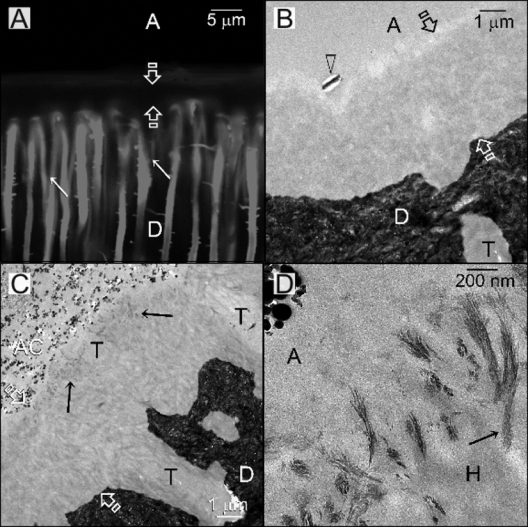
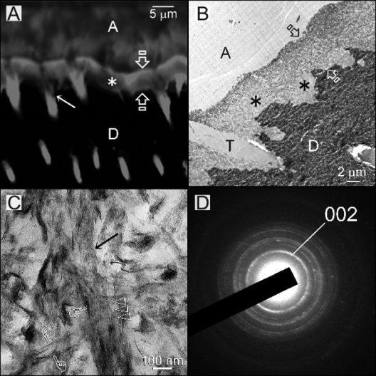
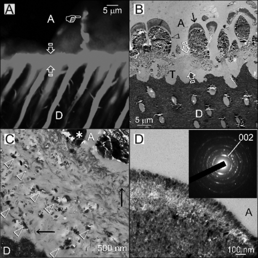
During mineralization, unbound water within the collagen matrix is replaced by apatite. This study tested the null hypothesis that there is no difference in the status of in vitro biomimetic remineralization of hybrid layers, regardless of their moisture contents. Acid-etched dentin was bonded with One-Step with ethanol-wet-bonding, water-wet-bonding, and water-overwet-bonding protocols. Composite-dentin slabs were subjected to remineralization for 1-4 months in a medium containing dual biomimetic analogs, with set Portland cement as the calcium source and characterized by transmission electron microscopy. Remineralization was either non-existent or restricted to the intrafibrillar mode in ethanol-wet-bonded specimens. Extensive intrafibrillar and interfibrillar remineralization was observed in water-wet-bonded specimens. Water-overwet specimens demonstrated partial remineralization of hybrid layers and precipitation of mineralized plates within water channels. The use of ethanol-wet-bonding substantiates that biomimetic remineralization is a progressive dehydration process that replaces residual water in hybrid layers with apatite crystallites.
Figures
Similar articles
-
The critical barrier to progress in dentine bonding with the etch-and-rinse technique.J Dent. 2011 Mar;39(3):238-48. doi: 10.1016/j.jdent.2010.12.009. Epub 2011 Jan 6. J Dent. 2011. PMID: 21215788 Free PMC article. Clinical Trial.
-
Biomimetic remineralization of resin-bonded acid-etched dentin.J Dent Res. 2009 Aug;88(8):719-24. doi: 10.1177/0022034509341826. J Dent Res. 2009. PMID: 19734458 Free PMC article. Clinical Trial.
-
Phosphoric acid esters cannot replace polyvinylphosphonic acid as phosphoprotein analogs in biomimetic remineralization of resin-bonded dentin.Dent Mater. 2009 Oct;25(10):1230-9. doi: 10.1016/j.dental.2009.05.001. Epub 2009 May 30. Dent Mater. 2009. PMID: 19481792 Free PMC article. Clinical Trial.
-
From dry bonding to water-wet bonding to ethanol-wet bonding. A review of the interactions between dentin matrix and solvated resins using a macromodel of the hybrid layer.Am J Dent. 2007 Feb;20(1):7-20. Am J Dent. 2007. PMID: 17380802 Review.
-
The use of bioactive particles and biomimetic analogues for increasing the longevity of resin-dentin interfaces: A literature review.Dent Mater J. 2020 Jan 31;39(1):62-68. doi: 10.4012/dmj.2019-293. Epub 2019 Nov 14. Dent Mater J. 2020. PMID: 31723068 Review.
Cited by
-
Limitations in bonding to dentin and experimental strategies to prevent bond degradation.J Dent Res. 2011 Aug;90(8):953-68. doi: 10.1177/0022034510391799. Epub 2011 Jan 10. J Dent Res. 2011. PMID: 21220360 Free PMC article. Review.
-
Evaluating the effect of poly (amidoamine) treated bioactive glass nanoparticle incorporated in universal adhesive on bonding to artificially induced caries affected dentin.BMC Oral Health. 2023 Oct 28;23(1):810. doi: 10.1186/s12903-023-03536-4. BMC Oral Health. 2023. PMID: 37898802 Free PMC article.
-
Therapeutic effects of novel resin bonding systems containing bioactive glasses on mineral-depleted areas within the bonded-dentine interface.J Mater Sci Mater Med. 2012 Jun;23(6):1521-32. doi: 10.1007/s10856-012-4606-6. Epub 2012 Apr 1. J Mater Sci Mater Med. 2012. PMID: 22466816
-
Biomimetic Mineralizing Agents Recover the Micro Tensile Bond Strength of Demineralized Dentin.Materials (Basel). 2018 Sep 14;11(9):1733. doi: 10.3390/ma11091733. Materials (Basel). 2018. PMID: 30223511 Free PMC article.
-
The critical barrier to progress in dentine bonding with the etch-and-rinse technique.J Dent. 2011 Mar;39(3):238-48. doi: 10.1016/j.jdent.2010.12.009. Epub 2011 Jan 6. J Dent. 2011. PMID: 21215788 Free PMC article. Clinical Trial.
References
-
- Becker TD, Agee KA, Joyce AP, Rueggeberg FA, Borke JL, Waller JL, et al. (2007). Infiltration/evaporation-induced shrinkage of demineralized dentin by solvated model adhesives. J Biomed Mater Res B Appl Biomater 80:156-165 - PubMed
-
- Bella J, Brodsky B, Berman HM. (1995). Hydration structure of a collagen peptide. Structure 3:893-906 - PubMed
-
- Bonar LC, Lees S, Mook HA. (1985). Neutron diffraction studies of collagen in fully mineralized bone. J Mol Biol 181:265-270 - PubMed
-
- Bowman SM, Gibson LJ, Hayes WC, McMahon TA. (1999). Results from demineralized bone creep tests suggest that collagen is responsible for the creep behavior of bone. J Biomech Eng 121:253-258 - PubMed
-
- Cameron IL, Short NJ, Fullerton GD. (2007). Verification of simple hydration/dehydration methods to characterize multiple water compartments on tendon type 1 collagen. Cell Biol Int 31:531-539 - PubMed
Publication types
MeSH terms
Substances
Grants and funding
LinkOut - more resources
Full Text Sources
Other Literature Sources
