. 2010 Apr;120(4):1097-110.
doi: 10.1172/JCI40979.

Hereditary spastic paraplegia proteins REEP1, spastin, and atlastin-1 coordinate microtubule interactions with the tubular ER network

Affiliations

Hereditary spastic paraplegia proteins REEP1, spastin, and atlastin-1 coordinate microtubule interactions with the tubular ER network

Seong H Park et al. J Clin Invest. 2010 Apr.

Abstract

Hereditary spastic paraplegias (HSPs; SPG1-45) are inherited neurological disorders characterized by lower extremity spastic weakness. More than half of HSP cases result from autosomal dominant mutations in atlastin-1 (also known as SPG3A), receptor expression enhancing protein 1 (REEP1; SPG31), or spastin (SPG4). The atlastin-1 GTPase interacts with spastin, a microtubule-severing ATPase, as well as with the DP1/Yop1p and reticulon families of ER-shaping proteins, and SPG3A caused by atlastin-1 mutations has been linked pathogenically to abnormal ER morphology. Here we investigated SPG31 by analyzing the distribution, interactions, and functions of REEP1. We determined that REEP1 is structurally related to the DP1/Yop1p family of ER-shaping proteins and localizes to the ER in cultured rat cerebral cortical neurons, where it colocalizes with spastin and atlastin-1. Upon overexpression in COS7 cells, REEP1 formed protein complexes with atlastin-1 and spastin within the tubular ER, and these interactions required hydrophobic hairpin domains in each of these proteins. REEP proteins were required for ER network formation in vitro, and REEP1 also bound microtubules and promoted ER alignment along the microtubule cytoskeleton in COS7 cells. A SPG31 mutant REEP1 lacking the C-terminal cytoplasmic region did not interact with microtubules and disrupted the ER network. These data indicate that the HSP proteins atlastin-1, spastin, and REEP1 interact within the tubularER membrane in corticospinal neurons to coordinate ER shaping and microtubule dynamics. Thus, defects in tubular ER shaping and network interactions with the microtubule cytoskeleton seem to be the predominant pathogenic mechanism of HSP.


Figures

Figure 1
Figure 1. REEP1 is in the DP1/Yop1p superfamily of ER-shaping proteins.
(A) REEP/DP1/Yop1p protein phylogeny. A ClustalW (version 1.4) tree with species name and GenBank protein accession numbers are shown. REEP1–6 are color coded. The scale bar denotes the number of substitutions per site. (B) Expression of REEP1. Untransfected HEK293 cells (Untrans), cells overexpressing REEP1, and rat brain and spinal cord (Sp cord) homogenates were immunoblotted for REEP1. An arrowhead indicates REEP1; an asterisk identifies modified REEP1 or a cross-reacting protein. MW standards (in kDa) are indicated throughout. (C) REEP1 antibody specificity. Cells overexpressing REEP1 were transfected with control or REEP1-specific siRNA and immunoblotted for REEP1. β-Tubulin was monitored as a loading control. (D) Oligomerization of REEP1. Extracts from REEP1-expressing cells were cross-linked with DSP and resolved by SDS-PAGE on nonreducing gels with or without DTT, which cleaves cross-links. (E) REEP1 membrane association. Homogenates (Homog) from REEP1-expressing cells were separated into soluble and membrane (Memb) fractions then immunoblotted for REEP1 or the cytoplasmic protein β-tubulin. (F) Alkaline membrane extraction. Lysed membranes from REEP1-expressing cells (Input) were alkaline extracted, and soluble and pellet fractions were immunoblotted for REEP1 and the soluble protein Grp78. (G) Detergent phase partitioning. Membranes from REEP1-expressing cells were partitioned with Triton X-114. Input membranes as well as detergent and aqueous phases were immunoblotted. (H) Cells overexpressing REEP1 and ΔN-REEP1 (lacking aa 1–20) were immunoblotted for REEP1. (I) Protease protection. Proteinase K (PK) was added to intact microsomes from REEP1-expressing cells with or without Triton X-100, and aliquots were immunoblotted. (J) Two possible topology models for REEP1–4. (K) Topology model for REEP5–6/Yop1p. Cyto, cytoplasm.
Figure 2
Figure 2. Anti–REEP1–4 antibodies inhibit ER network formation in vitro.
(A and B) Membranes from Xenopus eggs were incubated in the absence (A) or presence (B) of GTP for 1 hour, stained with octadecyl rhodamine, and visualized by confocal fluorescence microscopy. (CH) As in B, except membranes were preincubated for 1 hour with 1.1 μM control IgG (C), affinity-purified pan-atlastin antibodies (D), either of 2 different affinity-purified anti-REEP antibodies (E and F), anti-IP3R antibodies (G), or anti-TRAPα antibody (H) prior to addition of GTP. Scale bar: 20 μm.
Figure 3
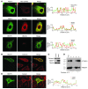
Figure 3. REEP1 expression increases ER alignment along microtubules.
(A) REEP1 is an ER protein. COS7 cells coexpressing REEP1 (green) and Mito-DsRed (red; top row), the ER protein RFP-Sec61β (red; middle row), or REEP1-expressing cells costained for the cis/medial-Golgi maker GM130 (red; bottom row) were imaged using confocal microscopy. Relative fluorescence intensities for the indicated linear regions in merged images were measured using Zeiss LSM510 software and are graphed. Note the high degree of REEP1 and Sec61β line scan overlap but lack of significant overlap between REEP1 and Mito-DsRed or GM130. (B) HeLa cells were transfected with REEP1 and costained for REEP1 (green) and endogenous calnexin (red). Two untransfected cells are present at the bottom of the image for comparison. (C) Aliquots of untransfected and REEP1-transfected cells were immunoblotted for calnexin, REEP1, and β-tubulin. (D) REEP1 interaction with microtubules. REEP1 levels are markedly increased in the pellet (P), along with tubulin, upon addition of paclitaxel and GTP to cell lysates. S, soluble fraction. (E) COS7 cells were transfected with REEP1 and costained for REEP1 (green) and endogenous β-tubulin (red). Note the high degree of line scan overlap. Scale bars: 10 μm.
Figure 4
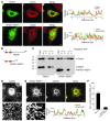
Figure 4. SPG31 REEP1 truncation mutant does not interact with microtubules and impairs ER reticularization.
(A and B) SPG31 mutant REEP1 (aa 1–112) does not colocalize with the microtubule cytoskeleton. (A) COS7 cells were transfected with WT (top row) or mutant (bottom row) REEP1 and costained for β-tubulin (red), with merged images and line scan plots shown. (B) A schematic diagram of the REEP1 forms. (C) Mutant REEP1 does not interact with microtubules. Microtubule-binding assays show clear enrichment of WT REEP1 in the pellet fraction of extracts from cells preincubated with paclitaxel and GTP. Mutant REEP1 remains in the soluble fraction even after addition of paclitaxel and GTP. MW standards (in kDa) are indicated. (D) Cells overexpressing GFP-Sec61β exhibit a typical ER network, as revealed with confocal fluorescence microscopy. (E) Cells coexpressing Sec61β and mutant REEP1 exhibit a disrupted ER, with loss of both 3-way junctions and tubular appearance, despite the extensive colocalization of REEP1 (green) and RFP-Sec61β (red), as shown in the merged image and line scan plot. (D and E) Boxed areas are enlarged in the panels below (original magnification, ×5.7). (F) SPG31 mutant REEP1 expression inhibits formation of 3-way ER junctions. Number of 3-way junctions in cells in similar 250 μm2 areas from D and E were counted (mean ± SD of 4 regions per trial, n = 3 trials for each condition; P < 0.001). Scale bars: 10 μm.
Figure 5
Figure 5. REEP1 C-terminal cytoplasmic domain is sufficient for microtubule interaction in vitro.
(A) GST fusion protein production. Aliquots of crude bacterial lysates and affinity-purified GST and GST-REEP1 (aa 113–201) fusion proteins are shown, with detection by Coomassie Brilliant Blue staining after SDS-PAGE. MW standards (in kDa) are indicated. (B) REEP1 interaction with polymerized microtubules. GST-REEP1 (aa 113–201) levels are markedly increased in the pellet, along with tubulin, upon addition of paclitaxel and GTP to purified microtubules, while GST remains in the soluble fraction. The lower 2 panels represent immunoblots from the same gel, with detection of GST-REEP1 (aa 113–201) using anti-REEP1 antibodies.
Figure 6
Figure 6. Endogenous REEP1 colocalizes with atlastin-1 and spastin in cerebral cortical neurons in culture.
(A) Membranes prepared from rat cortical neurons in primary culture were immunoblotted for REEP1. (B) REEP1 colocalizes with atlastin-1. Neurons were costained for REEP1 (green) and atlastin-1 (red). A merged image superimposed on the DIC image is shown, and boxed areas representing areas of enrichment and colocalization in growth cones (top panels), axonal varicosities (middle panels), and the axon shaft (bottom panels) are shown in enlargements to the right (original magnification, ×4.6). (C) REEP1 colocalizes with spastin. Neurons were costained for REEP1 (green) and spastin (red). The boxed area shows protein enrichment and colocalization in axonal varicosities, as shown in enlargements to the right (original magnification, ×2.5). Scale bars: 20 μm.
Figure 7
Figure 7. REEP1 interacts with atlastin-1 through intramembrane hydrophobic domains.
(A) The ER network in COS7 cells overexpressing REEP1 and atlastin-1 alone is morphologically distinct. Boxed areas are shown in enlargements to the right (original magnification, ×2.3). (B) Colocalization of overexpressed REEP1 and atlastin-1 in COS7 cells. Atlastin-1 (red) immunoreactive puncta are studded along REEP1-positive (green) tubules (top panels). The boxed area is enlarged in the panels directly below (original magnification, ×3.0). (C) Atlastin-1 and REEP1 coimmunoprecipitate. Cells were transfected with REEP1 and Myc–atlastin-1 or REEP1 alone, immunoprecipitated with either Myc-epitope or REEP1 antibodies, and immunoblotted with Myc-epitope and REEP1 antibodies. Input and control IgG IP lanes used extracts from cells coexpressing REEP1 and Myc–atlastin-1. (D) Domain mapping of atlastin-1 interaction with REEP1. Myc-tagged WT atlastin-1, atlastin-1 Ncyt (aa 1–447), or atlastin-1 TM (aa 449–558) were coexpressed with REEP1 and immunoprecipitated with anti-REEP1 antibodies. Immunoprecipitates were immunoblotted for Myc-epitope or REEP1. Arrowheads identify immunoprecipitated atlastin-1 proteins, and an asterisk identifies the IgG light chain. MW standards (in kDa) are to the left. (E) REEP1 (green) was coexpressed with Myc–atlastin-1 Ncyt or Myc–atlastin-1 TM (red) and identified in cells using confocal immunofluorescence microscopy. REEP1 colocalizes with atlastin-1 TM but not atlastin-1 Ncyt, as shown in the merged images and as quantitated in the line scan plots. Scale bars: 20 μm.
Figure 8
Figure 8. REEP1 interacts preferentially with the M1 isoform of spastin.
(A) A schematic diagram showing the domain organization of spastin isoforms generated through use of 2 different translation start codons. MIT, present in microtubule-interacting and transport proteins. (B) REEP1 (green) was coexpressed with Myc-tagged M87 (top; red) or M1 spastin (bottom; red) and visualized using confocal microscopy. Scale bars: 10 μm. (C) Alkaline extraction. Myc-tagged M1 spastin but not M87 spastin is present exclusively in the pellet fraction after alkaline extraction, as revealed by immunoblotting for Myc-epitope. MW standards (in kDa) are indicated throughout. (D) Detergent phase partitioning. Membranes from M1 and M87 spastin-expressing cells were partitioned with Triton X-114 (TX-114). Input membranes as well as aqueous (A) and detergent (D) phases were immunoblotted. Partitioning of the soluble protein Grp78 is shown for comparison. (E) Protease protection. Proteinase K was added to intact microsomes from Myc-tagged M1 spastin-expressing cells, with or without Triton X-100, and aliquots were immunoblotted for M1 spastin (Myc-epitope), Grp78, and calnexin. (F) M1 spastins Y52N and G15N create consensus N-linked glycosylation sites but are not glycosylated. WT and the indicated mutant M1 spastin proteins were expressed in COS7 cells and immunoblotted for Myc. (G) Model for REEP1 membrane topology. (H) M1 spastin and REEP1 coimmunoprecipitate. Cells were cotransfected with REEP1 and either Myc-tagged M1 or M87 spastin, then immunoprecipitated with anti-REEP1 antibodies and immunoblotted with anti–Myc-epitope antibodies. The control IgG IP lane used extracts from cells coexpressing REEP1 and M1 spastin.
Figure 9
Figure 9. Model for interactions among the HSP proteins in the tubular ER.
In this schematic diagram, atlastin-1 and M1 spastin interact directly with the ER-shaping proteins, including REEP1, as well as with one another (not shown). These interactions very likely occur through the hydrophobic hairpins of each of these proteins inserted into the membrane, though the first hydrophobic domain may exist in the ER membrane instead as a more traditional transmembrane segment (see Figure 1J). It is unclear whether the larger atlastin-1 hydrophobic segments completely span the membrane. REEP1 makes direct contact with the microtubule cytoskeleton through its C-terminal cytoplasmic domain. The M1 isoform of the spastin ATPase also binds to microtubules, through the MIT domain or a region adjacent to it, and is involved in microtubule severing, coupling changes in ER morphology with microtubule dynamics. GTP, GTP-binding domain; MTB, microtubule-binding domain.

References

    1. Reid E. Science in motion: common molecular pathological themes emerge in the hereditary spastic paraplegias. J Med Genet. 2003;40(2):81–86. doi: 10.1136/jmg.40.2.81. - DOI - PMC - PubMed
    1. Fink JK. Hereditary spastic paraplegia. Curr Neurol Neurosci Rep. 2006;6(1):65–76. doi: 10.1007/s11910-996-0011-1. - DOI - PubMed
    1. Soderblom C, Blackstone C. Traffic accidents: molecular genetic insights into the pathogenesis of the hereditary spastic paraplegias. Pharmacol Ther. 2006;109(1–2):42–56. doi: 10.1016/j.pharmthera.2005.06.001. - DOI - PubMed
    1. Züchner S. The genetics of hereditary spastic paraplegia and implications for drug therapy. Expert Opin Pharmacother. 2007;8(10):1433–1439. doi: 10.1517/14656566.8.10.1433. - DOI - PubMed
    1. Salinas S, Proukakis C, Crosby A, Warner TT. Hereditary spastic paraplegia: clinical features and pathogenetic mechanisms. Lancet Neurol. 2008;7(12):1127–1138. - PubMed

Publication types