. 2010 Apr;120(4):1310-23.
doi: 10.1172/JCI39987.

Induction of autophagy-dependent necroptosis is required for childhood acute lymphoblastic leukemia cells to overcome glucocorticoid resistance

Affiliations

Induction of autophagy-dependent necroptosis is required for childhood acute lymphoblastic leukemia cells to overcome glucocorticoid resistance

Laura Bonapace et al. J Clin Invest. 2010 Apr.

Abstract

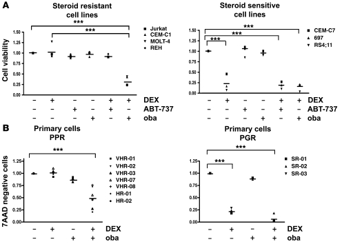
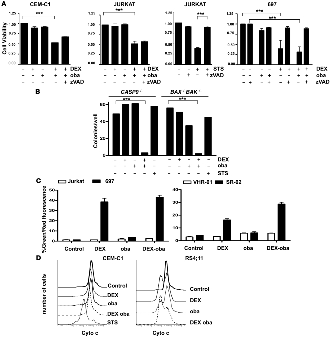
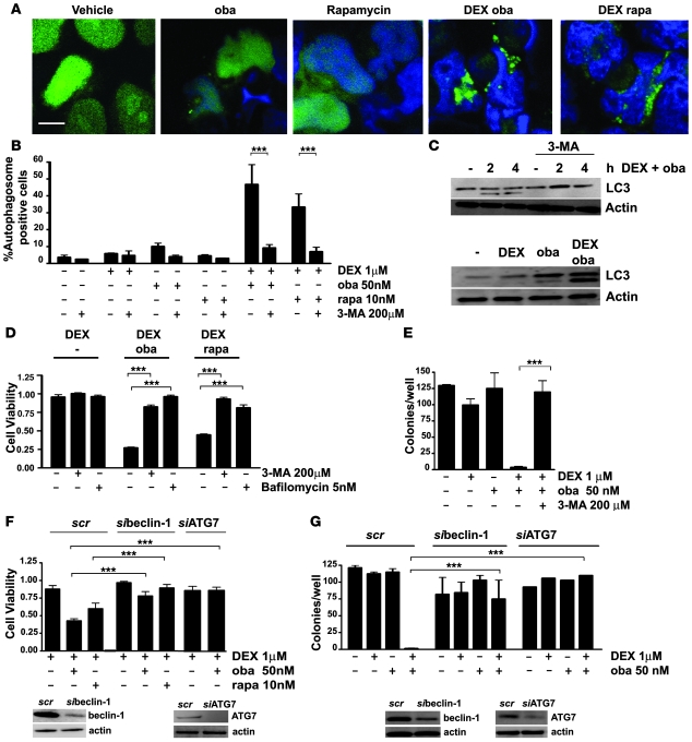
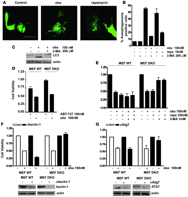
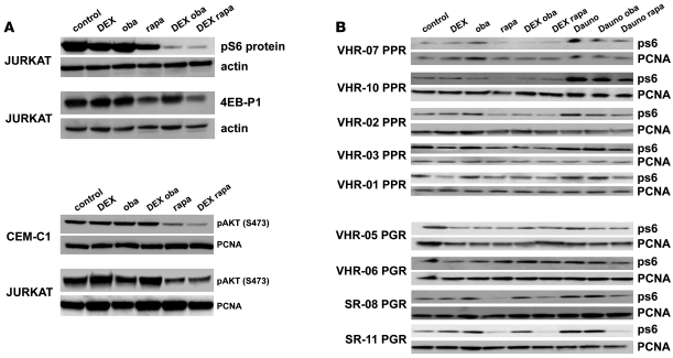
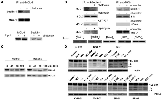
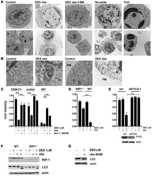
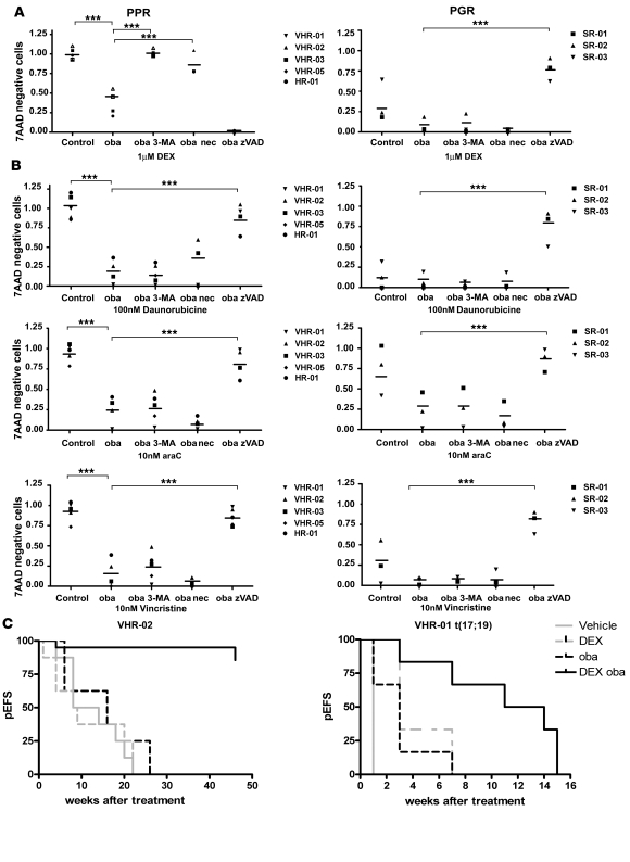
In vivo resistance to first-line chemotherapy, including to glucocorticoids, is a strong predictor of poor outcome in children with acute lymphoblastic leukemia (ALL). Modulation of cell death regulators represents an attractive strategy for subverting such drug resistance. Here we report complete resensitization of multidrug-resistant childhood ALL cells to glucocorticoids and other cytotoxic agents with subcytotoxic concentrations of obatoclax, a putative antagonist of BCL-2 family members. The reversal of glucocorticoid resistance occurred through rapid activation of autophagy-dependent necroptosis, which bypassed the block in mitochondrial apoptosis. This effect was associated with dissociation of the autophagy inducer beclin-1 from the antiapoptotic BCL-2 family member myeloid cell leukemia sequence 1 (MCL-1) and with a marked decrease in mammalian target of rapamycin (mTOR) activity. Consistent with a protective role for mTOR in glucocorticoid resistance in childhood ALL, combination of rapamycin with the glucocorticoid dexamethasone triggered autophagy-dependent cell death, with characteristic features of necroptosis. Execution of cell death, but not induction of autophagy, was strictly dependent on expression of receptor-interacting protein (RIP-1) kinase and cylindromatosis (turban tumor syndrome) (CYLD), two key regulators of necroptosis. Accordingly, both inhibition of RIP-1 and interference with CYLD restored glucocorticoid resistance completely. Together with evidence for a chemosensitizing activity of obatoclax in vivo, our data provide a compelling rationale for clinical translation of this pharmacological approach into treatments for patients with refractory ALL.


Figures

Figure 1
Figure 1. Obatoclax resensitizes GC-resistant ALL cells to dexamethasone.
Combination experiments were performed with subcytotoxic concentrations (10% IC50) of obatoclax (oba) or ABT-737 and 1 μM dexamethasone (DEX), and values were normalized to cells treated with vehicle control. (A) ALL cell lines were treated for 72 hours as indicated. Cell viability was determined by the MTT assay. (B) Primary ALL cells from 7 PPR patients with VHR-ALL and 3 prednisone-good-responder (PGR) patients were treated as indicated for 72 hours. Cell viability was assessed by flow cytometry using 7AAD. ***P < 0.05.
Figure 2
Figure 2. Obatoclax resensitizes GC-resistant ALL cells to dexamethasone without activation of mitochondrial apoptosis.
(A) ALL cells were treated as indicated for 48 hours, for controls STS or zVAD.fmk (80 nM) was used, and cell viability was assessed with the MTT assay. 697 cells served as GC-sensitive control. (B) Jurkat CASP9–/– and BAX–/–BAK–/– cells were treated for 72 hours as indicated, and clonogenic survival was assessed after incubation in methylcellulose for 7 days. (C) Percentages of cells with JC-1 monomers, corresponding to a loss of the mitochondrial potential, are shown for GC-resistant CEM-C1 and GC-sensitive CEM-C7 cells and in samples from PGR and PPR patients after treatment as indicated for 16 hours. (D) Cytochrome c release was induced in steroid-sensitive RS4;11 cells but not in the resistant CEM-C1 cell line upon treatment with dexamethasone or dexamethasone and obatoclax. STS was used as positive control. Cytochrome c release was detected by flow cytometry. ***P < 0.05.
Figure 3
Figure 3. Obatoclax induces autophagy in GC-resistant ALL cells.
(A) After transient transfection with GFP-LC3, Jurkat cells were treated for 4 hours as indicated. The characteristic punctuate staining pattern indicative of autophagosome formation was detected by confocal microscopy in cells treated with dexamethasone and obatoclax or rapamycin (rapa). Scale bar: 10 μm. (B) Quantitation of autophagosome-positive cells. The data represent mean ± SD of 2 independent experiments, counting 200 cells each. (C) Detection of endogenous LC3-II by Western blot analysis. Jurkat cells were treated with obatoclax and dexamethasone for the indicated time points in the presence or absence of 3-MA and after 24 hours of treatment as indicated. (D) Inhibition of autophagy by 3-MA or bafilomycin impaired sensitization to dexamethasone by 10 nM rapamycin or obatoclax (10% IC50). Cell viability was assessed by MTT. (E) Treatment with obatoclax and dexamethasone inhibited clonogenic survival of Jurkat cells. 3-MA rescued GC-resistant cells from cell death induced by combination treatment. Cells were treated for 72 hours with compounds, and clonogenic survival in methylcellulose was assessed after washing and incubation for 7 days. (F) Downregulation of beclin-1 or ATG7 using siRNA (si) impaired the resensitization of Jurkat cells to dexamethasone by obatoclax or rapamycin compared with scrambled (scr) controls. Cell viability was assessed by MTT. Efficiency of downregulation at 48 hours was analyzed by Western analysis. (G) Downregulation of beclin-1 and ATG7 in Jurkat cells protected cells from obatoclax- and dexamethasone-induced cell death in the clonogenic assay. ***P < 0.05.
Figure 4
Figure 4. Obatoclax and rapamycin induce autophagic cell death in Bax–/–Bak–/– MEFs.
(A) Bax–/–Bak–/– MEFs transiently expressing the autophagy marker GFP-LC3 were treated with vehicle, obatoclax (100 nM), or rapamycin (10 nM) for 4 hours. Autophagosome formation was monitored by confocal microscopy. Scale bar: 20 μm. (B) Quantitation of autophagosome-positive cells. The data represent mean ± SD of 2 independent experiments, counting 200 cells each. (C) Treatment of Bax–/–Bak–/– MEFs with obatoclax induced generation of endogenous LC3-II as detected by Western analysis, an effect which was blocked by 3-MA treatment. (D) WT or Bax–/–Bak–/– (DKO) MEFs were incubated for 48 hours with obatoclax or ABT-737, and cell viability assessed by the MTT assay. (E) Inhibition of autophagy by 3-MA rescued Bax–/–Bak–/– MEFs but not WT MEFs from cell death induced by 48 hour exposure to obatoclax or rapamycin. (F) Downregulation of beclin-1 rescued Bax–/–Bak–/– MEFs but not WT MEFs from cell death induced by obatoclax as evaluated by the MTT assay. Efficiency of downregulation was determined by Western blot analysis. (G) Downregulation of ATG7 rendered Bax–/–Bak–/– MEFs but not WT MEFs resistant to obatoclax treatment. Efficiency of downregulation was assessed by Western blotting.
Figure 5
Figure 5. Combination treatment with dexamethasone and obatoclax leads to inhibition of mTOR.
(A) Jurkat cells were treated for 6 hours as indicated, and phosphorylation of the mTOR targets, S6 protein and 4EB-P1, was assessed. Combination of dexamethasone with either obatoclax or rapamycin resulted in inhibition of mTOR activity (top panel). Rapamycin alone and in combination with dexamethasone, but not obatoclax alone or with dexamethasone, decreased the phosphorylation of AKT at Ser473 in Jurkat and CEM-C1 cells (bottom panel). (B) In primary cells from PPR patients with VHR-ALL, treatment with dexamethasone and obatoclax or rapamycin resulted in a decrease of S6 protein phosphorylation. In contrast, in cells from prednisone-good-responder patients, S6 protein was not dephosphorylated after combination treatment.
Figure 6
Figure 6. Obatoclax induces disruption of a complex of MCL-1 and beclin-1.
(A) FLAG-tagged beclin-1 was overexpressed in 293T cells, and MCL-1 immunoprecipitates were analyzed for the presence of beclin-1, with or without obatoclax treatment for 3 hours. (B) Jurkat cells were treated with 100 nM obatoclax, 30 nM ABT-737, or 10 nM rapamycin for 3 hours, and MCL-1 or beclin-1 immunoprecipitates were analyzed for the presence of proteins as indicated. Low-dose obatoclax disrupted the interaction between beclin-1 and MCL-1, while interactions between BCL2 and beclin-1 were unaffected. Likewise, ABT-737 or rapamycin did not affect complexes between beclin-1 and MCL-1 or BCL2. (C) MCL-1 levels were assessed after treatment with vehicle or the combination of dexamethasone and obatoclax at indicated time points, with or without cycloheximide (CHX). Western blot analyses revealed that MCL-1 levels were unaffected by combination treatment. (D) BIM induction was assessed by Western blot in steroid-resistant (Jurkat, VHR-01, and VHR-02) and steroid-sensitive (RS4;11, 697, SR-01, and SR-02) cell lines and primary cells treated with DEX (1 μM), obatoclax (100 nM), or the combination for 24 hours.
Figure 7
Figure 7. RIP-1 kinase activity is essential for obatoclax-mediated GC sensitization.
(A) Electron microscopy images reveal features of necroptotic cell death after treatment for 72 hours with obatoclax and dexamethasone. No condensed chromatin was detectable, while Trail treatment induces condensed apoptotic nuclei (top panel) (original magnification, ×7,100). Cells treated with obatoclax and dexamethasone exhibited disintegrated plasma membranes, which was recapitulated by Na-azide. Trail treatment leaved membranes intact (bottom panel) (original magnification, ×5,400). (B) A more detailed view of the same experiment. An autophagosome formation with characteristic double membrane structures was detected in the cytoplasm. The plasma membrane was disrupted in cells treated with obatoclax and dexamethasone. N, nucleus; C, cytoplasm; A, autophagosome; PM, plasma membrane (original magnification, ×15,000). (C) In steroid-resistant (CEM-C1 and Jurkat) and steroid-sensitive (697) cells treated for 72 hours with dexamethasone (1 μM) and obatoclax (10% IC50), with or without the necroptosis inhibitor nec-1 (300 nM), nec-1 restored steroid resistance as assessed by the MTT assay. (D) Jurkat RIP1–/– cells were less sensitive to the double treatment for 72 hours with dexamethasone (1 mM) and obatoclax (10% IC50) compared with WT Jurkat cells. Cell viability was assessed by the MTT assay. (E) Downregulation of CYLD rescued Jurkat cells from cell death induced by combination treatment for 72 hours with dexamethasone (1 μM) and obatoclax (10% IC50). Efficiency of downregulation was assessed by Western blot analysis after 48 hours. (F) LC3-II generation occurred in Jurkat RIP1–/– cells and in WT cells after 8 hours of treatment with dexamethasone and obatoclax. (G) Treatment with nec-1 did not inhibit LC3-II generation induced by treatment with obatoclax and dexamethasone in Jurkat cells. ***P < 0.05.
Figure 8
Figure 8. Obatoclax displays a strong chemosensitizing activity in multidrug-resistant primary ALL cells from poor risk patients.
(A) Primary ALL cells from 5 PPR patients and 3 prednisone-good-responder patients were cocultured with hTERT-immortalized bone marrow stroma cells and treated with either dexamethasone (1 μM) alone or in combination with obatoclax (10% IC50) in the presence or absence of 3-MA, nec-1, or zVAD.fmk for 72 hours. Cell viability was assessed by 7AAD staining and flow cytometry. Data are shown as mean ± SD of 2 independent experiments. In combination experiments, values were normalized to cells treated with compounds and/or inhibitors alone at indicated dose. (B) Primary ALL cells from 4 VHR, 1 high risk (HR), and 3 standard risk (SR) patients were cocultured with hTERT-immortalized bone marrow stroma cells and treated with either daunorubicin, vincristine, or cytarabine (araC) alone or in combination with obatoclax (10% IC50) in the presence or absence of 3-MA, nec-1, or zVAD.fmk for 72 hours. Cell viability was assessed by 7AAD staining and flow cytometry. Data are shown as mean ± SD of 2 independent experiments. ***P < 0.05 (A and B). (C) Percentage of event-free survival (pEFS) of NSG mice after xenotransplantation with primary cells from 2 PPR patients with VHR-ALL (VHR-02, n = 8; patient VHR-01, n = 6; Supplemental Table 2) and treatment for 3 weeks with either vehicle, dexamethasone, obatoclax, or the combination. P < 0.02 for dexamethasone and obatoclax versus vehicle, dexamethasone, or obatoclax alone for both xenograft experiments.

Similar articles

Cited by

References

    1. Pui CH, Evans WE. Treatment of acute lymphoblastic leukemia. N Engl J Med. 2006;354(2):166–178. doi: 10.1056/NEJMra052603. - DOI - PubMed
    1. Schrappe M. Evolution of BFM trials for childhood ALL. Ann Hematol. 2004;83(Suppl 1):S121–S123. - PubMed
    1. Schrauder A, et al. Prospective evaluation of MRD–Kinetics in 274 children with high-risk ALL treated in trial ALL–BFM 2000: Insights into development of resistance and impact on further refinement of treatment stratification strategies. Blood. 2007;110:585.
    1. Holleman A, et al. Gene-expression patterns in drug-resistant acute lymphoblastic leukemia cells and response to treatment. N Engl J Med. 2004;351(6):533–542. doi: 10.1056/NEJMoa033513. - DOI - PubMed
    1. Wei G, et al. Gene expression-based chemical genomics identifies rapamycin as a modulator of MCL1 and glucocorticoid resistance. Cancer Cell. 2006;10(4):331–342. doi: 10.1016/j.ccr.2006.09.006. - DOI - PubMed

Publication types

MeSH terms