Review
. 2010 Jul-Sep;4(3):447-57.
doi: 10.4161/cam.4.3.10771. Epub 2010 Jul 30.

Transitions between epithelial and mesenchymal states and the morphogenesis of the early mouse embryo

Affiliations
Review

Transitions between epithelial and mesenchymal states and the morphogenesis of the early mouse embryo

Anna Ferrer-Vaquer et al. Cell Adh Migr. 2010 Jul-Sep.

Abstract

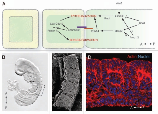
Multicellular organisms arise from the generation of different cell types and the organization of cells into tissues and organs. Cells of metazoa display two main phenotypes, the ancestral epithelial state and the recent mesenchymal derivative. Epithelial cells are usually stationary and reside in two-dimensional sheets. By contrast mesenchymal cells are loosely packed and can move to new positions, thereby providing a vehicle for cell rearrangement, dispersal and novel cell-cell interactions. Transitions between epithelial and mesenchymal states drive key morphogenetic events in the early vertebrate embryo, including gastrulation, germ layer formation and somitogenesis. The cell behaviors and molecular mechanisms promoting transitions between these two states in the early mouse embryo are discussed in this review.


Figures

Figure 1
Figure 1
Cell phenotypes in the early mouse embryo. (A) Gastrulating mouse embryo at embryonic day (E) 7.5. (C') Scanning electronic micrograph of a transverse section through a E7.5 mouse embryo showing the different cell phenotypes: columnar epithelium, mesenchyme and squamous epithelium. (B) Diagram of the embryonic part of a e7.5 mouse embryo. Dashed box outlines the three germ layers. (C) Scanning micrograph shown in (B and C') color-coded for the three germ layers.
Figure 2
Figure 2
Diagrammatic representation of MET, EMT, egression and ingression. (A) Mesenchymal cells undergo MeT to epithelialize, as for example during somitogenesis. Conversely, epithelial cells undergo eMT to assume mesenchymal characteristics. (B) in an ingression, cells undergo EMT and leave an epithelium, while in an egression cells undergo MET and join a preexisting epithelium.
Figure 3
Figure 3
EMT at the mouse primitive streak. (B') Scanning electronic micrograph showing a transverse section through a E7.5 mouse primitive streak. (A) Scheme of the embryonic part of a E7.5 mouse embryo. Dashed box outlines the primitive streak. (B) Scanning micrograph of (A) color-coded for the different germ layers. (C) Cells undergo an eMT event at the primitive streak. (1) First intercellular spaces appear between cells and (2) basal lamina breaks down. (3) Cells acquire a bottle shape, (4) round up as they travel through the streak, and (5) finally acquire a stellate morphology and migrate away from the streak. (D) Signaling pathways that regulate the different EMT steps at the murine primitive streak.
Figure 4
Figure 4
The two alternative models of endoderm morphogenesis in the mouse gastrula. (A) in the displacement model, the VE is dislodged to the extaembryonic region by the nascent DE as a coherent epithelium. In the dispersal model, the initially uniform VE epithelium (1) is interrupted by single egressing epiblast-derived cells at different sites (2). The VE-derived cells are further dispersed (3) until isolated as single cells in the gut epithelium (4). (B and C) Separating VE cells downregulate tight junction markers (bottom panels). GFp positive ve cells separating during the VE dispersal process downregulate the tight junction marker ZO-1 between their interfaces (white arrowhead), but keep tight junctions with surrounding DE cells intact. The two DE cells flanking the separating DE cells are possibly egressing and undergoing MET, thereby establishing tight junctions with the surrounding cells.
Figure 5
Figure 5
MET during somitogenesis. (A) Model showing the molecular pathways involved in intersomitic border formation and somite epithelialization in amniotes. Anterior to the left. (B) Somites (dashed box) appear in a rostro-caudal fashion at the dorsal side of a ten somite-stage mouse embryo. (C) Scanning electronic micrograph of 4 somitic blocks from a ten somite-stage mouse embryo. (D) Confocal image showing the mesenchymal core surrounded by epithelium in an epithelized somite. Phalloidin (red) labels the actin cytoskeleton and Hoechst (blue), the nuclei.

Similar articles

Cited by

References

    1. Bertet C, Sulak L, Lecuit T. Myosin-dependent junction remodelling controls planar cell intercalation and axis elongation. Nature. 2004;429:1–3. - PubMed
    1. Blankenship JT, Backovic ST, Sanny JS, Weitz O, Zallen JA. Multicellular rosette formation links planar cell polarity to tissue morphogenesis. Dev Cell. 2006;11:459–470. - PubMed
    1. Pilot F, Lecuit T. Compartmentalized morphogenesis in epithelia: from cell to tissue shape. Dev Dyn. 2005;232:685–694. - PubMed
    1. Cano A, Pérez-Moreno M, Rodrigo I, Locascio A, Blanco M, del Barrio M, et al. The transcription factor snail controls epithelial-mesenchymal transitions by repressing E-cadhrin expression. Nat Cell Biol. 2000;2:76–83. - PubMed
    1. Hay E. Organization and fine structure of epithelium and mesenchyme in the developing chick embryo. Baltimore: Williams and Wilkins; 1968.

Publication types

LinkOut - more resources