. 2010 Jun;35(7):1510-9.
doi: 10.1038/npp.2010.20. Epub 2010 Mar 3.

Impact of lipid raft integrity on 5-HT3 receptor function and its modulation by antidepressants

Affiliations

Impact of lipid raft integrity on 5-HT3 receptor function and its modulation by antidepressants

Caroline Nothdurfter et al. Neuropsychopharmacology. 2010 Jun.

Abstract

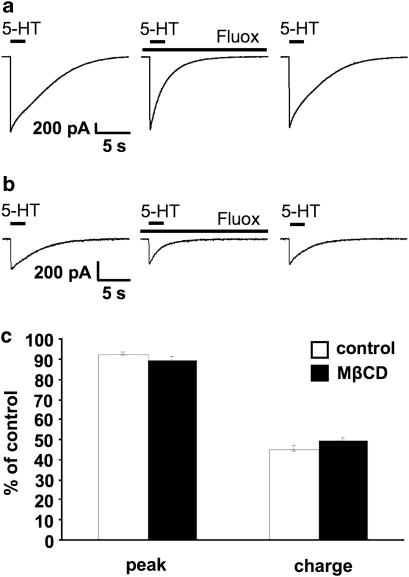
Because of the biochemical colocalization of the 5-HT(3) receptor and antidepressants within raft-like domains and their antagonistic effects at this ligand-gated ion channel, we investigated the impact of lipid raft integrity for 5-HT(3) receptor function and its modulation by antidepressants. Treatment with methyl-beta-cyclodextrine (MbetaCD) markedly reduced membrane cholesterol levels and caused a more diffuse membrane distribution of the lipid raft marker protein flotillin-1 indicating lipid raft impairment. Both amplitude and charge of serotonin evoked cation currents were diminished following cholesterol depletion by either MbetaCD or simvastatin (Sim), whereas the functional antagonistic properties of the antidepressants desipramine (DMI) and fluoxetine (Fluox) at the 5-HT(3) receptor were retained. Although both the 5-HT(3) receptor and flotillin-1 were predominantly found in raft-like domains in western blots following sucrose density gradient centrifugation, immunocytochemistry revealed only a coincidental degree of colocalization of these two proteins. These findings and the persistence of the antagonistic effects of DMI and Fluox against 5-HT(3) receptors after lipid raft impairment indicate that their modulatory effects are likely mediated through non-raft 5-HT(3) receptors, which are not sufficiently detected by means of sucrose density gradient centrifugation. In conclusion, lipid raft integrity appears to be important for 5-HT(3) receptor function in general, whereas it is not a prerequisite for the antagonistic properties of antidepressants such as DMI and Fluox at this ligand-gated ion channel.


Figures

Figure 1
Figure 1
Treatment with MβCD decreases serotonin-evoked cation currents in N1E-115 cells. (a) Effect of MβCD under cholesterol-depleting conditions. Cation currents were recorded in a whole-cell voltage-clamp configuration. 30 μM 5-HT was applied for 2 s. The left panel shows representative recordings of an untreated cell (control; without FCS) and of a cell pretreated with 0.5 mM MβCD for 12 h (MβCD). The left panel shows currents of a representative experiment, the right panel shows the same recording normalized to control. (b) Effect of MβCD in the presence of FCS avoiding cholesterol depletion. Cation currents were recorded in a whole-cell voltage-clamp configuration. 30 μM 5-HT was applied for 2 s. The left panel shows representative recordings of an untreated cell in the presence of FCS (+FCS) and of a cell pretreated with 0.5 mM MβCD for 12 h (+FCS +MβCD). Cells were kept in DMEM with FCS for 12 h both during control and during MβCD incubation before the recordings. The left panel shows currents of a representative experiment, the right panel shows the same recording normalized to the untreated cell+FCS.
Figure 2
Figure 2
Treatment with MβCD impairs lipid raft integrity in N1E-115 cells. (a) Reduction of cholesterol content of membranes following MβCD treatment. One proportion of living cells remained untreated (control), the other proportion was treated with 0.5 mM MβCD for 12 h. Cells were then resuspended in high-salt HEPES buffer, homogenized and sonificated. After a two-step centrifugation, the second supernatant enriched with membranes was quantified for cholesterol and protein content. Results represent the mean±SEM of three independent experiments. The asterisk indicates a significant difference from control experiments (*P<0.05). (b) Treatment with MβCD induces a more diffuse distribution of flotillin-1. After fixation and permeabilization cells were immunostained with an antibody against flotillin-1 (red spots), cell nuclei were stained with DAPI (blue). Immunofluorescence was detected by confocal microscopy at a 60-fold magnification. Before immunocytochemistry one proportion of living cells was pretreated with 0.5 mM MβCD for 12 h, whereas the other proportion remained untreated (control). Representative images of three independent experiments are shown. (c) The more diffuse distribution of flotillin-1 in immunocytochemistry following MβCD treatment is not due to an altered expression of flotillin-1. Equal amounts of membrane homogenates were analyzed by SDS-PAGE. Replicate gels were immunoblotted with an antibody against flotillin-1. The blot shows a representative experiment of three independent experiments with untreated cells (control) and cells pretreated with 0.5 mM MβCD for 12 h.
Figure 3
Figure 3
The modulation of serotonin evoked currents by DMI is retained after MβCD or Sim treatment of N1E-115 cells. (a) Antagonistic effect of DMI at the 5-HT3 receptor. Cation currents were recorded in a whole-cell voltage-clamp configuration. 30 μM 5-HT were applied for 2 s in the absence or presence of 1 μM DMI. The upper bars indicate the presence of 5-HT, the lower bar indicates the presence of DMI. Representative recordings before and after application of DMI and subsequent washout are shown. (b) Antagonistic effect of DMI after MβCD treatment. Cation currents were recorded in a whole-cell voltage-clamp configuration. 30 μM 5-HT were applied for 2 s in the absence or presence of 1 μM DMI. The upper bars indicate the presence of 5-HT, the lower bar indicates the presence of DMI. Representative recordings before and after application of DMI and subsequent washout are shown. Cells were treated with 0.5 mM MβCD for 12 h before the recordings. (c) Antagonistic effect of DMI after Sim treatment. Cation currents were recorded in a whole-cell voltage-clamp configuration. 30 μM 5-HT were applied for 2 s in the absence or presence of 1 μM DMI. The upper bars indicate the presence of 5-HT, the lower bar indicates the presence of DMI. Representative recordings before and after application of DMI and subsequent washout are shown. Cells were treated with 0.5 μM Sim for 24 h before the recordings. (d) Peak amplitude and charge of controls (white bars) and after pretreatment with 0.5 mM MβCD for 12 h (black bars) or 0.5 μM Sim for 24 h (grey bars) in the presence of DMI of six independent experiments in relation to values in the absence of DMI, which are set as 100%. Data are shown as mean±SEM % of control. DMI reduces peak amplitude and charge to a similar degree in all three experimental conditions.
Figure 4
Figure 4
The modulation of serotonin evoked currents by Fluox is retained after MβCD treatment of N1E-115 cells. (a) Antagonistic effect of Fluox at the 5-HT3 receptor. Cation currents were recorded in a whole-cell voltage-clamp configuration. 30 μM 5-HT were applied for 2 s in the absence or presence of 3 μM Fluox. The upper bars indicate the presence of 5-HT, the lower bar indicates the presence of Fluox. Representative recordings before and after application of Fluox and subsequent washout are shown. (b) Antagonistic effect of Fluox after MβCD treatment. Cation currents were recorded in a whole-cell voltage-clamp configuration. 30 μM 5-HT was applied for 2 s in the absence or presence of 3 μM Fluox. The upper bars indicate the presence of 5-HT, the lower bar indicates the presence of Fluox. Representative recordings before and after application of Fluox and subsequent washout are shown. Cells were treated with 0.5 mM MβCD for 12 h before the recordings. (c) Peak amplitude and charge of controls (white bars) and after pretreatment with 0.5 mM MβCD for 12 h (black bars) in the presence of Fluox of six independent experiments in relation to values in the absence of Fluox, which are set as 100%. Data are shown as mean±SEM % of control. Fluox reduces peak amplitude and charge to a similar degree in both experimental conditions.
Figure 5
Figure 5
Treatment with high concentrations of MβCD shifts flotillin-1, caveolin, and the 5-HT3 receptor to the high buoyant density fractions (HBD) in sucrose gradients of HEK 293 cells stably expressing the human 5-HT3A receptor. Living cells were treated with MβCD for 12 h at different concentrations (0.5 and 7.5 mM); one proportion of the cells remained untreated (control). Cells were then homogenized, sonificated in high-salt HEPES buffer, and fractionated by sucrose gradient density centrifugation. Thereafter, 10 fractions were collected from the top to the bottom of the gradient and analyzed by SDS-PAGE. Replicate gels were immunoblotted with antibodies against flotillin-1 (a), caveolin (b) and the 5-HT3 receptor (c). The blots shown are representative gradients out of three independent experiments. LBD: low buoyant density fractions; HBD: high buoyant density fractions.
Figure 6
Figure 6
Low degree of colocalization of the 5-HT3 receptor with the lipid raft marker protein flotillin-1 in N1E-115 cells using immunofluorescence. (a) Cells were fixed, permeabilized, and immunostained with an antibody against flotillin-1 (red spots) and against the 5-HT3 receptor (green spots). Cell nuclei were stained in blue with DAPI. Immunofluorescence was recorded at a 60-fold magnification and a 6-fold zoom. Colocalization of flotillin-1 and the 5-HT3 receptor is indicated by the superposition of red and green fluorescence in the form of yellow spots (merge). Representative images out of five independent experiments are shown. (b) Magnification of the merged images from the yellow box in (a) without DAPI. Intensity profiles along the blue lines of the red channel (flotillin-1) and the green channel (5-HT3 receptor) show only a low degree of colocalization of flotillin-1 and the 5-HT3 receptor (white arrow). (c) The proportion of double-labeled spots within one cell (flotillin-1 labeled (flottilin-1+) and 5-HT3 receptor labeled (5-HT3+) was quantified in relation to the total number of flotillin-1+ spots to assess the relative percentage of raft-associated 5-HT3 receptors (raft). This was compared with the proportion of 5-HT3 receptor+/flotillin-1 non-labeled (flotillin-1) spots in relation to total 5-HT3 receptor+ spots to quantify the relative percentage of non-raft-associated 5-HT3 receptors (non-raft). The relative percentage of raft-5-HT3 receptors was significantly lower (21.9±2.3%) in comparison with that of non-raft-5-HT3 receptors (78.2±3.6%) as indicated by one-way ANOVA analysis of the results from five independent experiments (F(1)=161.96; ***p⩽0.001). There was no difference in immunofluorescence patterns between cells kept in DMEM supplemented with 10% FCS compared with cells kept in serum-free DMEM for 12 h before immunocytochemistry (data not shown).

Similar articles

Cited by

References

    1. Allen JA, Halverson-Tamboli RA, Rasenick MM. Lipid raft microdomains and neurotransmitter signalling. Nat Rev Neurosci. 2007;8:128–140. - PubMed
    1. Bari M, Oddi S, De Simone C, Spagnolo P, Gasperi V, Battista N, et al. Type-1 cannabinoid receptors colocalize with caveolin-1 in neuronal cells. Neuropharmacology. 2008;54:45–50. - PMC - PubMed
    1. Barnes NM, Sharp T. A review of central 5-HT receptors and their function. Neuropharmacology. 1999;38:1083–1152. - PubMed
    1. Barrantes FJ. Cholesterol effects on nicotinic acetylcholine receptor. J Neurochem. 2007;103 (Suppl 1:72–80. - PubMed
    1. Baumann P, Gaillard JM, Perey M, Justafre JC, Le P. Relationships between brain concentrations of desipramine and paradoxical sleep inhibition in the rat. J Neural Transm. 1983;56:105–116. - PubMed

Publication types

MeSH terms