

Comparative Study
doi: 10.1038/gt.2010.22.
Epub 2010 Mar 4.
Cutaneous vaccination using microneedles coated with hepatitis C DNA vaccine
Affiliations
- PMID: 20200562
- PMCID: PMC2914565
- DOI: 10.1038/gt.2010.22
Item in Clipboard
Comparative Study
Cutaneous vaccination using microneedles coated with hepatitis C DNA vaccine
Gene Ther.
2010 Jun.
Abstract
The skin is potentially an excellent organ for vaccine delivery because of accessibility and the presence of immune cells. However, no simple and inexpensive cutaneous vaccination method is available. Micron-scale needles coated with DNA were tested as a simple, inexpensive device for skin delivery. Vaccination with a plasmid encoding hepatitis C virus nonstructural 3/4A protein using microneedles effectively primed specific cytotoxic T lymphocytes (CTLs). Importantly, the minimally invasive microneedles were as efficient in priming CTLs as more complicated or invasive delivery techniques, such as gene gun and hypodermic needles. Thus, microneedles may offer a promising technology for DNA vaccination.
Figures
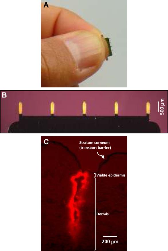
Microneedles were cut from stainless steel sheets using an infrared laser and electropolished as described before . (A) Photograph of a representative microneedle row held in a human hand. (B) Microneedle row with five microneedles uniformly coated with vitamin B2 as a model compound. Because plasmid coatings were difficult to visualize, uniformity of coatings was assessed by coating microneedles with a model colored compound, vitamin B2 (riboflavin-5'-phosphate sodium salt dihydrate) (Fisher Scientific, Fair Lawn, NJ, USA). The aqueous dip-coating solution contained 1% (w/v) carboxymethylcellulose sodium salt (CMC, low viscosity, USP grade, CarboMer, San Diego, CA, USA), 0.5% (w/v) Lutrol F-68 NF (BASF, Mt. Olive, NJ, USA) and 20 mg/ml vitamin B2 . Microneedles were coated using a custom dip-coating device and air-dried for >24h . For immunization, microneedles were coated using the same formulation, but by replacing vitamin B2 with 5 mg/ml codon optimized NS3/4A plasmid DNA. To determine the amount of DNA coated on microneedles, DNA concentration was measured by a validated technique using UV absorbance at 260 nm in a solution prepared by vortexing coated microneedles for 1 min in 1ml deionized water. (C) Histological section of porcine cadaver skin after inserting sulforhodamine-coated microneedle. To histologically characterize insertion of microneedles into skin, microneedles were coated with 0.1% (w/v) sulforhodamine (Molecular Probes, Eugene, OR, USA) and inserted into porcine cadaver skin for 1 min.
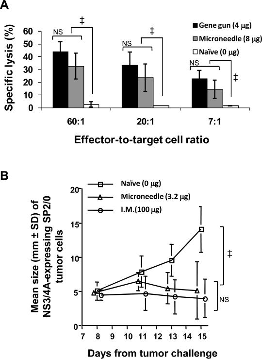
Groups of 4-8 week old female C57BL/6 or Balb/c mice (Charles River, Uppsala, Sweden) were immunized cutaneously with microneedles or gene gun, or intramusculary using hypodermic needles.For microneedle-based immunization two or five rows per mouse were used to control the dose. Each microneedle row was coated with 1.6 μg DNA. DNA-coated microneedle rows were manually inserted into trimmed abdominal or back skin and held for 1 min to allow dissolution of coated DNA into skin. For gene gun-based immunization mice were immunized by gene gun (Bio-Rad Laboratories, Hercules, CA, USA) at a dose of 4 μg/mouse as previously described . Plasmid DNA was linked to 1-μm diameter gold particles for gene gun-based immunizations according to protocols supplied by the manufacturer. For intramuscular immunization mice were injected with 100 μg DNA in the tibialis anterior using a hypodermic needle. Un-treated (naive) mice were used as a negative control. All experimental protocols were approved by the ethical committee for animal research at Karolinska Institutet. (A) C57B1/6 mice immunized once with codon-optimized NS3/4A DNA using either microneedles (1.6 μg per row × 5 microneedle rows per mouse = 8 μg dose; n=4 mice; gray bars), gene gun (4 μg dose; n=4 mice; black bars) or no immunization (n=2 mice; white bars) and euthanized after two weeks. As described previously ,, cells harvested from the spleen (effector cells, 2.5×10⁷ cells per mouse) were restimulated with a NS3 H-2Db-specific peptide and 2.5×10⁷ irradiated naïve C57B1/6 spleenocytes cells. After five days, 5×10³ RMA-S cells pulsed with NS3 H-2Db-specific peptide and labeled with 51Cr were used as target cells. The specific cell lysis of target cells was then measured at different effector-to-target cell ratios by measuring 51Cr released from lysed target cells. (B) Balb/C mice were either immunized intramuscularly (100 μg; n=5 mice),cutaneously with microneedles (1.6 μg per row × 2 microneedle rows per mouse = 3.2 μg dose; n=5 mice) or were not immunized (n=2 mice). Presence of in vivo functional CTLs was determined using a tumor challenge model . Two weeks after immunization, mice were subcutaneously injected with 1×10⁶ SP2/0 myeloma cells stably transfected with NS3/4A. Tumor growth was then monitored daily through skin by recording mean tumor size (thickness of skin flap at tumor injection site) for 14 days and compared to growth of the same tumor cell line in non-vaccinated mice. Mean tumor sizes were compared by analysis of variance (ANOVA, α=0.05). Error bars represent SD; Symbol ‡ represents p<0.05; NS means not significant.
Similar articles
-
In vivo clearance of hepatitis C virus nonstructural 3/4A-expressing hepatocytes by DNA vaccine-primed cytotoxic T lymphocytes.J Infect Dis. 2005 Dec 15;192(12):2112-6. doi: 10.1086/498218. Epub 2005 Nov 4. J Infect Dis. 2005. PMID: 16288375
-
Codon optimization and mRNA amplification effectively enhances the immunogenicity of the hepatitis C virus nonstructural 3/4A gene.Gene Ther. 2004 Mar;11(6):522-33. doi: 10.1038/sj.gt.3302184. Gene Ther. 2004. PMID: 14999224
-
Effective humoral immune response from a H1N1 DNA vaccine delivered to the skin by microneedles coated with PLGA-based cationic nanoparticles.J Control Release. 2017 Nov 10;265:66-74. doi: 10.1016/j.jconrel.2017.04.027. Epub 2017 Apr 20. J Control Release. 2017. PMID: 28434892
-
Microneedle-based vaccines.Curr Top Microbiol Immunol. 2009;333:369-93. doi: 10.1007/978-3-540-92165-3_18. Curr Top Microbiol Immunol. 2009. PMID: 19768415 Free PMC article. Review.
-
Revealing the potential of DNA-based vaccination: lessons learned from the hepatitis B virus surface antigen.Biol Chem. 2001 Apr;382(4):543-52. doi: 10.1515/BC.2001.068. Biol Chem. 2001. PMID: 11405219 Review.
Cited by
-
Microneedle-based drug and vaccine delivery via nanoporous microneedle arrays.Drug Deliv Transl Res. 2015 Aug;5(4):397-406. doi: 10.1007/s13346-015-0238-y. Drug Deliv Transl Res. 2015. PMID: 26044672 Free PMC article. Review.
-
Progress in microneedle array patch (MAP) for vaccine delivery.Hum Vaccin Immunother. 2021 Jan 2;17(1):316-327. doi: 10.1080/21645515.2020.1767997. Epub 2020 Jul 15. Hum Vaccin Immunother. 2021. PMID: 32667239 Free PMC article.
-
Simple and customizable method for fabrication of high-aspect ratio microneedle molds using low-cost 3D printing.Microsyst Nanoeng. 2019 Sep 9;5:42. doi: 10.1038/s41378-019-0088-8. eCollection 2019. Microsyst Nanoeng. 2019. PMID: 31645996 Free PMC article.
-
Microneedle-based intradermal delivery of stabilized dengue virus.Bioeng Transl Med. 2019 Feb 25;4(2):e10127. doi: 10.1002/btm2.10127. eCollection 2019 May. Bioeng Transl Med. 2019. PMID: 31249877 Free PMC article.
-
Coating solid dispersions on microneedles via a molten dip-coating method: development and in vitro evaluation for transdermal delivery of a water-insoluble drug.J Pharm Sci. 2014 Nov;103(11):3621-3630. doi: 10.1002/jps.24159. Epub 2014 Sep 11. J Pharm Sci. 2014. PMID: 25213295 Free PMC article.
References
-
- Garmory HS, Perkins SD, Phillpotts RJ, Titball RW. DNA vaccines for biodefence. Adv Drug Deliver Rev. 2005;57:1343–1361. - PubMed
-
- Donnelly J, Berry K, Ulmer JB. Technical and regulatory hurdles for DNA vaccines. Int J Parasitol. 2003;33:457–467. - PubMed
-
- Nicolas JF, Guy B. Intradermal, epidermal and transcutaneous vaccination: from immunology to clinical practice. Expert Rev Vaccines. 2008;7:1201–1214. - PubMed
-
- Prausnitz MR, Mikszta JA, Cormier M, Andrianov AK. Microneedle-based vaccines. In: Compans RW, Orenstein WA, editors. Current Topics in Microbiology & Immunology: Vaccines for Pandemic Influenza. Springer-Verlag; Berlin/Heidelberg, Germany: 2009.
-
- Matriano JA, Cormier M, Johnson J, Young WA, Buttery M, Nyam K, et al. Macroflux microprojection array patch technology: a new and efficient approach for intracutaneous immunization. Pharm Res. 2002;19:63–70. - PubMed
Publication types
MeSH terms
Substances
Grants and funding
LinkOut - more resources
Full Text Sources
Other Literature Sources
