. 2010 Jan 22;20(2):247-258.
doi: 10.1002/adfm.200901172.

Time Controlled Protein Release from Layer-by-Layer Assembled Multilayer Functionalized Agarose Hydrogels

Affiliations

Time Controlled Protein Release from Layer-by-Layer Assembled Multilayer Functionalized Agarose Hydrogels

Sumit Mehrotra et al. Adv Funct Mater. .

Abstract

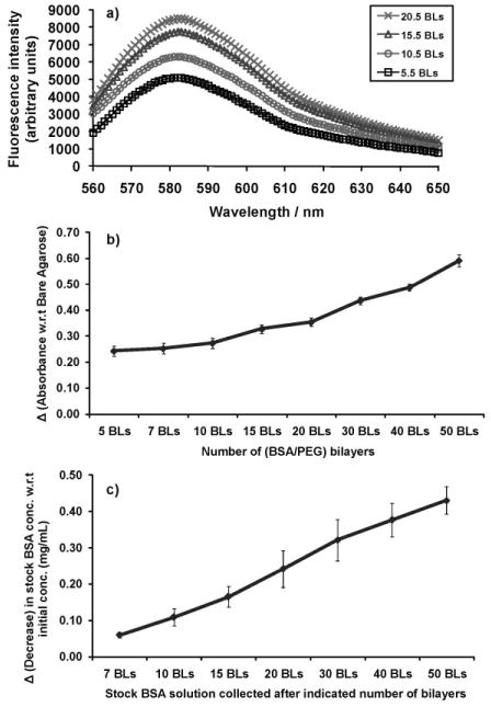
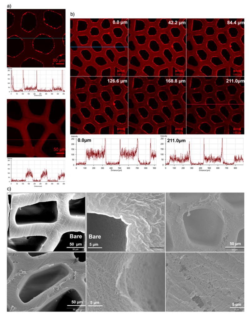
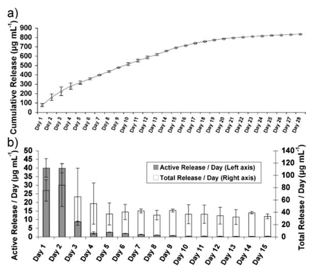
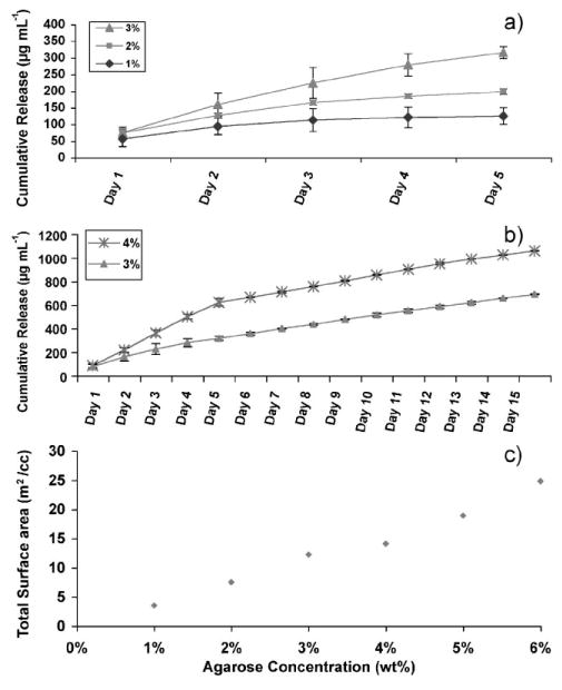
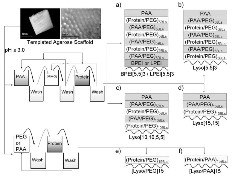
Axons of the adult central nervous system exhibit an extremely limited ability to regenerate after spinal cord injury. Experimentally generated patterns of axon growth are typically disorganized and randomly oriented. Support of linear axonal growth into spinal cord lesion sites has been demonstrated using arrays of uniaxial channels, templated with agarose hydrogel, and containing genetically engineered cells that secrete brain-derived neurotrophic factor (BDNF). However, immobilizing neurotrophic factors secreting cells within a scaffold is relatively cumbersome, and alternative strategies are needed to provide sustained release of BDNF from templated agarose scaffolds. Existing methods of loading the drug or protein into hydrogels cannot provide sustained release from templated agarose hydrogels. Alternatively, here it is shown that pH-responsive H-bonded poly(ethylene glycol)(PEG)/poly(acrylic acid)(PAA)/protein hybrid layer-by-layer (LbL) thin films, when prepared over agarose, provided sustained release of protein under physiological conditions for more than four weeks. Lysozyme, a protein similar in size and isoelectric point to BDNF, is released from the multilayers on the agarose and is biologically active during the earlier time points, with decreasing activity at later time points. This is the first demonstration of month-long sustained protein release from an agarose hydrogel, whereby the drug/protein is loaded separately from the agarose hydrogel fabrication process.


Figures

Figure 1
Figure 1
a) Fluorescence intensity measurements of TRITC conjugated to amine terminated PEG (PEG-Amine-TRITC) showed increased adsorption of PEG onto the agarose structure during the LbL deposition of BSA/(PEG-Amine-TRITC) multilayers on agarose hydrogel. b) UV/Vis absorbance measurements at 280 nm showed increased adsorption of bovine serum albumin (BSA) protein onto the agarose structure during the LbL deposition of BSA/(PEG) multilayers. The absorbance values are shown with respect to the absorbance of bare agarose, that is, the difference between the absorbance of LbL coated agarose and the absorbance of bare (non-coated) agarose. Five precursor bilayers of (PAA/PEG) with LPEI as the LbL initiating polymer were built over agarose in each case. BLs denote the number of bilayers. c) Corresponding decrease in lysozyme concentration in the bulk solution from the initial concentration as a function of the number of bilayers.
Figure 2
Figure 2
Confocal and scanning electron microscopy images showing the formation of LbL assembled multilayer thin films on agarose hydrogel substrate. a) Confocal microscopy images and associated fluorescence intensity profiles of a section below the top surface of agarose scaffolds coated with 30 bilayers of PAA and PEG followed by three bilayers of PEG and TRITC conjugated BSA (top image), and of agarose scaffolds coated with only 3 bilayers by PEG and TRITC conjugated BSA (bottom image). b) Confocal microscopy acquired z-section images of agarose scaffolds coated with 30 bilayers of PAA and PEG followed by three bilayers of PEG and TRITC conjugated BSA. Fluorescence intensity profiles of the topmost section (top panel: leftmost image) and a middle section (bottom panel: rightmost image) are shown. c) Scanning electron microscopy images of the scaffolds LbL coated with 30 bilayers of PAA and PEG. “Bare” denotes the non-coated scaffolds. BPEI was used as the LbL initiating polyelectrolyte for PAA/PEG assemblies.
Figure 3
Figure 3
a) Cumulative lysozyme release up to 4 weeks triggered by physiological pH, from LbL multilayer (as shown in Scheme 1a; BPEI initiating) coated 3% agarose hydrogel. b) Comparison between the total and enzymatically active lysozyme released per day, corresponding to the protein released in Figure 3a. Active concentrations were calculated from a standard curve obtained from pure lysozyme used to determine the degree of lysis of Micrococcus lysodeikticus by lysozyme.
Figure 4
Figure 4
a,b) Cumulative lysozyme release over time triggered by physiological pH from agarose hydrogel of varying concentrations, coated with LbL multilayer assembly (as shown in Scheme 1a; BPEI initiating). a) Comparison between 1%, 2%, and 3% agarose. b) Comparison between 3% and 4% agarose. c) Total surface area per unit volume of pure agarose hydrogel as a function of agarose concentration determined by BET.
Figure 5
Figure 5
The effect of LbL initiating polymer on lysozyme release triggered by physiological pH, from LbL coated 3% agarose hydrogel. BPEI, LPEI or lysozyme was used as the LbL initiating polymer as shown in Scheme 1a and b.
Figure 6
Figure 6
Cumulative lysozyme release from LbL-coated 3% agarose hydrogel, triggered by physiological pH, a) with varying stacking order of the polymers within a multilayer but the same number of cumulative bilayers (as shown in Scheme 1b–d) and b) with two-component assembly of lysozyme and PAA, two-component assembly of lysozyme and PEG, and three component assembly of PAA, PEG and lysozyme (as shown in Scheme 1f, e, and b respectively).
Figure 7
Figure 7
SEM images of H-bonded films composed of 25 bilayers of 10 kDa PEG and 15 kDa PAA formed on a planar substrate and exposed to deionized water (DI) (pH 5.6–6.3) for the time durations indicated. Top panel: SEM images of films after fabrication. Middle panel: SEM images of films after immersion in DI water for five days. Bottom panel: SEM images of films after immersion in DI water for ten days. Same spot on the films before and after degradation were imaged for comparative analysis. Columns 1, 2, and 3 show three different spots on the film.
Figure 8
Figure 8
a) Phase contrast microscopy images demonstrating the cytophobicity of the 30.5 bilayers of PAA/PEG multilayers over time (days). Top panel: NIH-3T3 fibroblasts cells on TCPS plates coated with 30.5 bilayers of PAA/PEG. Bottom panel: Fibroblasts on bare TCPS plates (control). b) Cytotoxicity levels of fibroblast cells after 4 days of culturing on (PAA/PEG)5PAA multilayers built onto TCPS (denoted here as 5.5BLs), (PAA/PEG)30PAA multilayers built onto TCPS (denoted here as 30.5 BLs), and bare TCPS plates. Serum in the cell culture medium contains a small amount of LDH, which is shown as “background media”. The absorbance values corresponds to the amount of lactate dehydrogenase (LDH) released into the culture supernatant.
Scheme 1
Scheme 1
Scheme diagram showing the three- and two-components LbL assembly fabricated onto native agarose as the substrate. Templated agarose scaffolds (as shown in this scheme) were used to characterize film growth, and agarose-filled TCPS plates were used to characterize protein releases. Three component assemblies consisted of PAA, PEG and protein as the multilayer constituents, and two component assemblies consisted of PEG or PAA and protein as the multilayer constituents. BPEI, LPEI, or protein (lysozyme, denoted as Lyso) was used as the LbL initiating polymer in the different cases shown in (a–f). Curved lines in (a–f) represent the agarose, and BLs indicates the bilayers.

Similar articles

Cited by

References

    1. Stokols S, Sakamoto J, Breckon C, Holt T, Weiss J, Tuszynski MH. Tissue Eng. 2006;12:2777. - PubMed
    1. Tuszynski MH, Gabriel K, Gage FH, Suhr S, Meyer S, Rosetti A. Exp Neurol. 1996;137:157. - PubMed
    1. Blesch A, Tuszynski MH. J Comp Neurol. 2003;467:403. - PubMed
    1. Bradbury EJ, Khemani S, King VR, Priestley JV, McMahon SB. Eur J Neurosci. 1999;11:3873. - PubMed
    1. Menei P, Montero-Menei C, Whittemore SR, Bunge RP, Bunge MB. Eur J Neurosci. 1998;10:607. - PubMed

LinkOut - more resources