Conscious and nonconscious memory effects are temporally dissociable
- PMID: 20200601
- PMCID: PMC2830725
- DOI: 10.1080/17588920903474263
Conscious and nonconscious memory effects are temporally dissociable
Abstract
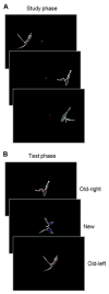
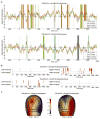
Intentional (explicit) retrieval can reactivate sensory cortex, which is widely assumed to reflect conscious processing. In the present study, we used an explicit visual memory event-related potential paradigm to investigate whether such retrieval related sensory activity could be separated into conscious and nonconscious components. During study, abstract shapes were presented in the left or right visual field. During test, old and new shapes were presented centrally and participants classified each shape as "old-left", "old-right", or "new". Conscious activity was isolated by comparing accurate memory for shape and location (old-hits) with forgotten shapes (old-misses), and nonconscious activity was isolated by comparing old-left-misses with old-right-misses and vice versa. Conscious visual sensory activity had a late temporal onset (after 800 ms) while nonconscious visual sensory activity had an early temporal onset (before 800 ms). These results suggest explicit memory related sensory activity reflects both conscious and nonconscious processes that are temporally dissociable.
Figures


Similar articles
-
Conscious processing during retrieval can occur in early and late visual regions.Neuropsychologia. 2013 Feb;51(3):482-7. doi: 10.1016/j.neuropsychologia.2012.11.020. Epub 2012 Nov 21. Neuropsychologia. 2013. PMID: 23178958 Free PMC article.
-
Nonconscious memory for motion activates MT+.Neuroreport. 2014 Nov 12;25(16):1326-30. doi: 10.1097/WNR.0000000000000270. Neuroreport. 2014. PMID: 25275641
-
The sensory timecourses associated with conscious visual item memory and source memory.Behav Brain Res. 2015 Sep 1;290:143-51. doi: 10.1016/j.bbr.2015.04.045. Epub 2015 May 5. Behav Brain Res. 2015. PMID: 25952962
-
The porous boundaries between explicit and implicit memory: behavioral and neural evidence.Ann N Y Acad Sci. 2011 Apr;1224:174-190. doi: 10.1111/j.1749-6632.2010.05946.x. Ann N Y Acad Sci. 2011. PMID: 21486300 Review.
-
Is implicit memory associated with the hippocampus?Cogn Neurosci. 2024 Apr;15(2):56-70. doi: 10.1080/17588928.2024.2315816. Epub 2024 Feb 18. Cogn Neurosci. 2024. PMID: 38368598
Cited by
-
"Chasing the first high": memory sampling in drug choice.Neuropsychopharmacology. 2020 May;45(6):907-915. doi: 10.1038/s41386-019-0594-2. Epub 2020 Jan 2. Neuropsychopharmacology. 2020. PMID: 31896119 Free PMC article. Review.
-
From Memory to Attitude: The Neurocognitive Process beyond Euthanasia Acceptance.PLoS One. 2016 Apr 18;11(4):e0153910. doi: 10.1371/journal.pone.0153910. eCollection 2016. PLoS One. 2016. PMID: 27088244 Free PMC article.
-
Involuntary and voluntary memory retrieval relies on distinct neural representations and oscillatory processes.PLoS Biol. 2025 Aug 19;23(8):e3003258. doi: 10.1371/journal.pbio.3003258. eCollection 2025 Aug. PLoS Biol. 2025. PMID: 40828871 Free PMC article.
-
Conscious processing during retrieval can occur in early and late visual regions.Neuropsychologia. 2013 Feb;51(3):482-7. doi: 10.1016/j.neuropsychologia.2012.11.020. Epub 2012 Nov 21. Neuropsychologia. 2013. PMID: 23178958 Free PMC article.
-
Episodic Memory Retrieval Functionally Relies on Very Rapid Reactivation of Sensory Information.J Neurosci. 2016 Jan 6;36(1):251-60. doi: 10.1523/JNEUROSCI.2101-15.2016. J Neurosci. 2016. PMID: 26740665 Free PMC article.
References
-
- Benjamini Y, Hochberg Y. Controlling the false discovery rate: A practical and powerful approach to multiple testing. Journal of the Royal Statistical Society: Series B. 1995;57:289–300.
-
- Clark VP, Fan S, Hillyard SA. Identification of early visual evoked potential generators by retinotopic and topographic analysis. Human Brain Mapping. 1995;2:170–187.
-
- Curran T, Schacter DL, Johnson MK, Spinks R. Brain potentials reflect behavioral differences in true and false recognition. Journal of Cognitive Neuroscience. 2001;13:201–216. - PubMed
Grants and funding
LinkOut - more resources
Full Text Sources
