. 2010 Feb;18(2):90-8.
doi: 10.1007/BF03091744.

Cardiac PET-CT: advanced hybrid imaging for the detection of coronary artery disease

Affiliations

Cardiac PET-CT: advanced hybrid imaging for the detection of coronary artery disease

P Knaapen et al. Neth Heart J. 2010 Feb.

Abstract

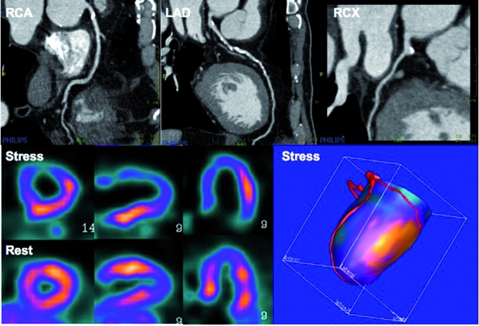
Hybrid imaging of positron emission tomography (PET) together with computed tomography (CT) is rapidly emerging. In cardiology, this new advanced hybrid imaging modality allows quantification of cardiac perfusion in combination with assessment of coronary anatomy within a single scanning session of less than 45 minutes. The near-simultaneous anatomical evaluation of coronary arteries using CT and corresponding functional status using PET provides a wealth of complementary information in patients who are being evaluated for (suspected) coronary artery disease, and could help guide clinical patient management in a novel manner. Clinical experience gained with this recently introduced advanced hybrid imaging tool, however, is still limited and its implementation into daily clinical practice remains largely unchartered territory. This review discusses principles of perfusion PET, its diagnostic accuracy, and potential clinical applications of cardiac PET-CT in patients with ischaemic heart disease. (Neth Heart J 2010;18:90-8.).

Keywords: Coronary Artery Disease; Positron-Emission Tomography; Tomography; X-Ray Computed.


Figures

Figure 1
Figure 1
Physics of positron emission, annihilation, and coincidence detection. Adapted from Paolo Camici.
Figure 2
Figure 2
A) Transaxial image at the thoracic level obtained a few minutes after injection of H215O. Note that the myocardium cannot be distinguished from the ventricular blood pool cavities as a result of free diffusion of tracer between blood and myocardial tissue, rendering the images unsuitable for diagnostic purposes. B) Through a mathematical process (i.e. factor or cluster analysis) the blood pool can be identified and subtracted from the H215O image, yielding an image of tracer activity distribution in the myocardial wall. These images can be used to qualitative grade tracer uptake. C) Parametric image of the same patient as in panel A and B. Perfusion is calculated at the voxel level through a modelling procedure based on the dynamic flux of tracer between arterial blood and myocardial tissue. Note the enhanced visual quality of the image in comparison with panel B. Moreover, for the parametric image perfusion is given in absolute terms (ml · min−1 · g−1) in the accompanying colour-coded legend.
Figure 3
Figure 3
A 52-year-old diabetic patient with atypical chest pain. The coronary arteries were severely calcified with a calcium score of more than 3500 (Agatston), therefore no CTCA was performed given its low diagnostic value in this type of patient due to blooming artefacts. PET perfusion with oxygen-15-labelled water displayed homogeneous perfusion both during rest and pharmacologically induced vasodilation (adenosine). Quantitative stress perfusion, however, was severely blunted (1.30 ml • min–1 • g–1, population-based normal value 3.54±1.01 ml • min–1 • g–1). Such a uniform distribution pattern would have resulted in a false-negative SPECT examination. The patient was referred for conventional angiography which revealed three-vessel coronary artery disease, confirming the findings of the hybrid PET-CT study.
Figure 4
Figure 4
Potential cardiac PET-CT scanning protocol. After an initial CT scout to delineate the field of view, pharmacological stress is induced by infusion of adenosine intravenously. Maximum vasodilation reaches a steady state after two minutes and the radionuclide is injected followed immediately by a dynamic PET frame sequence (approximately 5 minutes, which varies slightly for different PET tracers). Directly after the PET sequence, a CT attenuation scan (CTAC) is acquired after which the infusion of adenosine is terminated. Generally, stress imaging is performed early in the protocol to allow the heart rate to return to baseline level in order to reach a low heart rate during the CT coronary angiography (CTCA). After sufficient time to allow for radiodecay of the tracer, the rest PET sequence and a separate CTAC are acquired. Subsequently, a calcium score is performed (CTCAC) followed by CTCA during infusion of a contrast agent. During CTCAC and CTCA, a low and steady heart rate of less than 65 beats/min must be ensured. If necessary, intravenous β-blockers are administered to achieve this goal. The length of the protocol depends on the PET tracer used. It can be completed in less than 45 minutes with H215O and 82Rubidium, and in less than one hour with 13NH3.
Figure 5
Figure 5
Cardiac PET-CT images of a patient who is being evaluated for coronary artery disease. CTCA images display calcified lesions in the proximal RCA and throughout the proximal course of the LAD. PET images display a large reversible perfusion defect in the anterior wall, coinciding with the LAD territory on the fusion images, whereas the RCA perfusion territory has a normal hyperaemic response. This type of imaging therefore facilitates target vessel revascularisation in case of multivessel disease. Please note that the entire acquisition protocol is completed in less than 45 minutes.

Similar articles

Cited by

References

    1. Knaapen P, Lubberink M. Cardiac positron emission tomography: myocardial perfusion and metabolism in clinical practice. Clin Res Cardiol. 2008;97:791–6. - PubMed
    1. Kaufmann PA, Camici PG. Myocardial blood flow measurement by PET: technical aspects and clinical applications. J Nucl Med. 2005;46:75–88. - PubMed
    1. Di Carli MF, Hachamovitch R. New technology for noninvasive evaluation of coronary artery disease. Circulation. 2007;115:1464–80. - PubMed
    1. Le Guludec D, Lautamaki R, Knuuti J, Bax JJ, Bengel FM. Present and future of clinical cardiovascular PET imaging in Europe a position statement by the European Council of Nuclear Cardiology (ECNC). Eur J Nucl Med Mol Imaging. 2008;35:1709–24. - PubMed
    1. Schroeder S, Achenbach S, Bengel F, Burgstahler C, Cademartiri F, de Feyter P, et al. Cardiac computed tomography: indications, applications, limitations, and training requirements: report of a Writing Group deployed by the Working Group Nuclear Cardiology and Cardiac CT of the European Society of Cardiology and the European Council of Nuclear Cardiology. Eur Heart J. 2008;29:531–56. - PubMed

LinkOut - more resources