

Endoluminal abdominal aortic aneurysm repair: the latest advances in prevention of distal endograft migration and type 1 endoleak
- PMID: 20200623
- PMCID: PMC2829788
Endoluminal abdominal aortic aneurysm repair: the latest advances in prevention of distal endograft migration and type 1 endoleak
Abstract
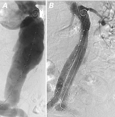
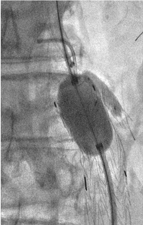
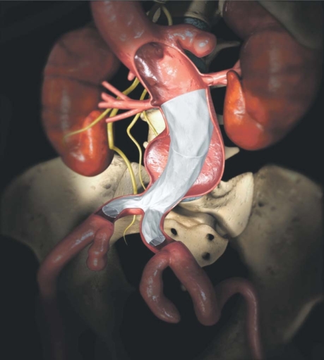
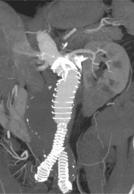
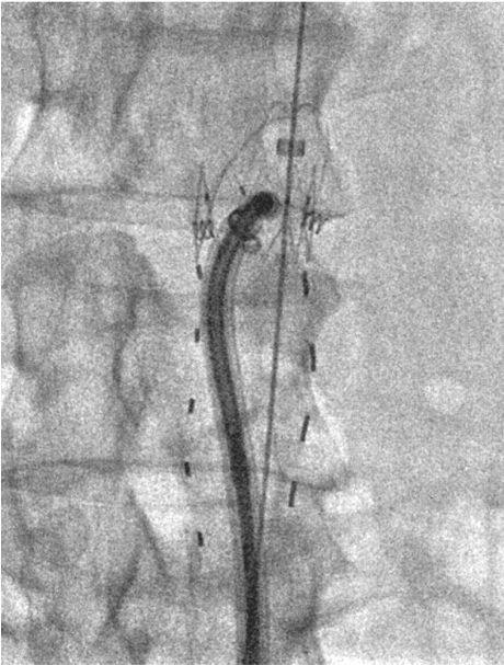
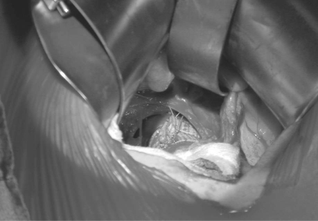
Endovascular abdominal aortic aneurysm repair (EVAR) is an attractive alternative to open surgical repair. Distal endograft migration and type 1 endoleak are recognized to be the 2 main complications of EVAR. First-generation endografts had a stronger propensity for distal migration, modular component separation, thrombosis, and loss of structural integrity. Substantial progress has been made in recent years with 2nd- and 3rd-generation devices to prevent these complications. Some of the most common predictors of endograft failure are angulated and short infrarenal necks, large-diameter necks, and thrombus in the aneurysmal sac. The purpose of this study is to describe and review our experience in using innovative techniques and a newer generation of endografts to prevent distal migration and type 1 endoleak in patients with challenging infrarenal neck anatomy. The use of these innovative EVAR techniques and the new generation of endografts in patients with challenging infrarenal neck anatomy has yielded encouraging procedural and intermediate-term results.
Keywords: Aneurysm, dissecting; aorta, abdominal; aortic aneurysm; aortic diseases; blood vessel prosthesis implantation; foreign-body migration; prosthesis design; stents.
Figures
Similar articles
-
Analysis of EndoAnchors for endovascular aneurysm repair by indications for use.J Vasc Surg. 2014 Dec;60(6):1460-7.e1. doi: 10.1016/j.jvs.2014.08.089. Epub 2014 Oct 3. J Vasc Surg. 2014. PMID: 25284629
-
Aortic Curvature Is a Predictor of Late Type Ia Endoleak and Migration After Endovascular Aneurysm Repair.J Endovasc Ther. 2017 Jun;24(3):411-417. doi: 10.1177/1526602817700378. Epub 2017 Mar 28. J Endovasc Ther. 2017. PMID: 28349777
-
Midterm outcome of EndoAnchors for the prevention of endoleak and stent-graft migration in patients with challenging proximal aortic neck anatomy.J Endovasc Ther. 2015 Apr;22(2):163-70. doi: 10.1177/1526602815574685. J Endovasc Ther. 2015. PMID: 25809354
-
Endograft accommodation on the aortic bifurcation: an overview of anatomical fixation and implications for long-term stent-graft stability.J Endovasc Ther. 2011 Aug;18(4):462-70. doi: 10.1583/11-3411.1. J Endovasc Ther. 2011. PMID: 21861731 Review.
-
Long-term follow-up after endovascular aneurysm repair: is ultrasound alone enough?Perspect Vasc Surg Endovasc Ther. 2010 Sep;22(3):145-51. doi: 10.1177/1531003510382664. Perspect Vasc Surg Endovasc Ther. 2010. PMID: 21098495 Review.
Cited by
-
Surgery for late type Ia/IIIb endoleak from a fabric tear and stent fracture of AFX2 stent graft.J Vasc Surg Cases Innov Tech. 2022 Jul 5;8(3):458-461. doi: 10.1016/j.jvscit.2022.06.005. eCollection 2022 Sep. J Vasc Surg Cases Innov Tech. 2022. PMID: 36016704 Free PMC article.
-
Secondary interventions following endovascular repair of abdominal aortic aneurysm.Gen Thorac Cardiovasc Surg. 2014 Feb;62(2):87-94. doi: 10.1007/s11748-013-0333-2. Epub 2013 Oct 22. Gen Thorac Cardiovasc Surg. 2014. PMID: 24146136 Review.
References
-
- Parodi JC, Palmaz JC, Barone HD. Transfemoral intraluminal graft implantation for abdominal aortic aneurysms. Ann Vasc Surg 1991;5(6):491–9. - PubMed
-
- Fulton JJ, Farber MA, Sanchez LA, Godshall CJ, Marston WA, Mendes R, et al. Effect of challenging neck anatomy on mid-term migration rates in AneuRx endografts. J Vasc Surg 2006;44(5):932–7. - PubMed
-
- Albertini J, Kalliafas S, Travis S, Yusuf SW, Macierewicz JA, Whitaker SC, et al. Anatomical risk factors for proximal perigraft endoleak and graft migration following endovascular repair of abdominal aortic aneurysms. Eur J Vasc Endovasc Surg 2000;19(3):308–12. - PubMed
-
- Harris PL, Vallabhaneni SR, Desgranges P, Becquemin JP, van Marrewijk C, Laheij RJ. Incidence and risk factors of late rupture, conversion, and death after endovascular repair of infrarenal aortic aneurysms: the EUROSTAR experience. European collaborators on stent/graft techniques for aortic aneurysm repair. J Vasc Surg 2000;32(4):739–49. - PubMed
-
- Chaikof EL, Blankensteijn JD, Harris PL, White GH, Zarins CK, Bernhard VM, et al. Reporting standards for endovascular aortic aneurysm repair. J Vasc Surg 2002;35(5):1048–60. - PubMed
Publication types
MeSH terms
LinkOut - more resources
Full Text Sources
Medical