

Non-gadolinium-enhanced 3-dimensional magnetic resonance angiography for the evaluation of thoracic aortic disease: a preliminary experience
- PMID: 20200628
- PMCID: PMC2829812
Non-gadolinium-enhanced 3-dimensional magnetic resonance angiography for the evaluation of thoracic aortic disease: a preliminary experience
Abstract
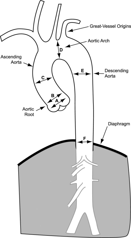
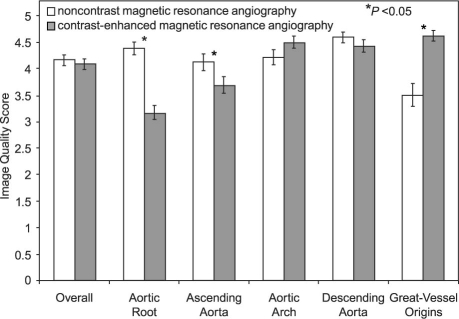
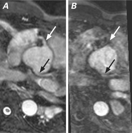
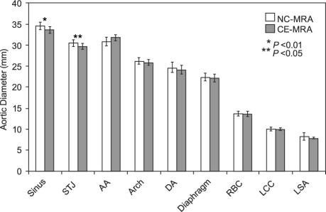
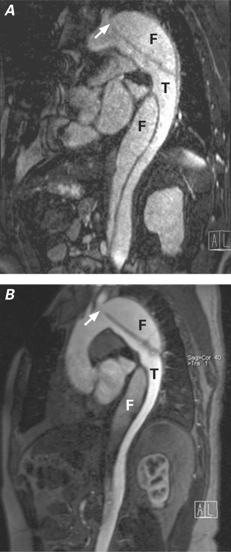
We compared image quality and diagnostic accuracy of a noncontrast 3-dimensional magnetic resonance angiography (NC-MRA) technique (balanced steady-state free-precession sequence) to contrast-enhanced MRA (CE-MRA) for evaluation of thoracic aortic disease.The CE-MRA provides 3-dimensional high-resolution images of the thoracic aorta that are important in the evaluation of patients with aortic disease. However, recent concerns with the potential nephrotoxic effects of gadolinium contrast medium limit the application of CE-MRA for patients who have significant renal insufficiency.Twenty-one patients (mean age, 51 yr; 18 men) who underwent NC-MRA and CE-MRA for evaluation of thoracic aortic disease were retrospectively identified. Data sets were reviewed by 2 readers who were blinded to the patients' information. The thoracic aorta was divided into 5 segments. Image quality and reader confidence for diagnosis of aortic pathology were rated on 5-point scales. The Wilcoxon matched-pairs signed rank test and the Student t test were used for comparisons.The NC-MRA identified all pathologic findings with 100% diagnostic accuracy and similar reader confidence, when compared with CE-MRA. Although overall image quality was not significantly different, superior image quality was observed at the aortic root (4.4 +/- 0.8 vs 3.2 +/- 0.9, P <0.0005) and ascending aorta (4.1 +/- 1 vs 3.7 +/- 0.9, P=0.05) respectively.In conclusion, NC-MRA is a useful alternative for evaluation and follow-up of thoracic aortic disease, especially for patients with poor intravenous access or contraindications to gadolinium use.
Keywords: Aneurysm, dissecting/diagnosis; aorta, thoracic/pathology; aortic aneurysm, thoracic/diagnosis; aortic diseases/diagnosis/radiography; artifacts; contrast media/toxici-ty; gadolinium/diagnostic use/toxicity; magnetic resonance angiography; retrospective studies.
Figures
References
-
- Krinsky GA, Rofsky NM, DeCorato DR, Weinreb JC, Earls JP, Flyer MA, et al. Thoracic aorta: comparison of gadolinium-enhanced three-dimensional MR angiography with conventional MR imaging. Radiology 1997;202(1):183–93. - PubMed
-
- Neimatallah MA, Ho VB, Dong Q, Williams D, Patel S, Song JH, Prince MR. Gadolinium-enhanced 3D magnetic resonance angiography of the thoracic vessels. J Magn Reson Imaging 1999;10(5):758–70. - PubMed
-
- Loewe C, Schillinger M, Haumer M, Loewe-Grgurin M, Lammer J, Thurnher S, et al. MRA versus DSA in the assessment of occlusive disease in the aortic arch vessels: accuracy in detecting the severity, number, and length of stenoses. J Endovasc Ther 2004;11(2):152–60. - PubMed
-
- Othersen JB, Maize JC, Woolson RF, Budisavljevic MN. Nephrogenic systemic fibrosis after exposure to gadolinium in patients with renal failure. Nephrol Dial Transplant 2007;22 (11):3179–85. - PubMed
-
- Broome DR, Girguis MS, Baron PW, Cottrell AC, Kjellin I, Kirk GA. Gadodiamide-associated nephrogenic systemic fibrosis: why radiologists should be concerned. AJR Am J Roentgenol 2007;188(2):586–92. - PubMed
Publication types
MeSH terms
Substances
LinkOut - more resources
Full Text Sources