Extrinsic compression of the left coronary ostium by the pulmonary trunk: management in a case of Eisenmenger syndrome
- PMID: 20200637
- PMCID: PMC2829817
Extrinsic compression of the left coronary ostium by the pulmonary trunk: management in a case of Eisenmenger syndrome
Abstract
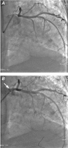
Extrinsic compression of the left main coronary artery by a massively dilated pulmonary artery in patients who have severe pulmonary hypertension can lead to significant myocardial ischemia. A 58-year-old man with a large patent ductus arteriosus and Eisenmenger syndrome presented with angina at rest and worsening heart failure of 3 months' duration. The new symptoms were recognized to be secondary to extrinsic compression of the left main coronary artery ostium by a dilated main pulmonary artery and were successfully relieved by the placement of a metallic stent in the affected segment of the left main coronary artery. Multislice computed tomographic imaging after 6 months showed stent patency and the intimate relation of the stented vessel to the dilated main pulmonary trunk. We discuss diagnostic and management issues pertaining to this uncommon clinical entity.
Keywords: Angina pectoris/etiology; Eisenmenger complex/ complications; angioplasty, transluminal, percutaneous coronary; constriction, patho-logic/etiology; coronary stenosis/etiology; dilatation, pathologic/complications; ductus arteriosus, patent; hypertension, pulmonary/complications; stents; tomography, X-ray computed.
Figures


Similar articles
-
Percutaneous Coronary Intervention for a Patient with Left Main Coronary Compression Syndrome.Intern Med. 2018 May 15;57(10):1421-1424. doi: 10.2169/internalmedicine.9534-17. Epub 2018 Jan 11. Intern Med. 2018. PMID: 29321426 Free PMC article. Review.
-
A case of extrinsic compression of the left main coronary artery secondary to pulmonary artery dilatation.J Korean Med Sci. 2013 Oct;28(10):1543-8. doi: 10.3346/jkms.2013.28.10.1543. Epub 2013 Sep 25. J Korean Med Sci. 2013. PMID: 24133364 Free PMC article.
-
Acute coronary syndrome due to extrinsic compression of the left main coronary artery in a patient with severe pulmonary hypertension: successful treatment with percutaneous coronary intervention.Cardiovasc Revasc Med. 2008 Jan-Mar;9(1):47-51. doi: 10.1016/j.carrev.2007.07.003. Cardiovasc Revasc Med. 2008. PMID: 18206638
-
Percutaneous revascularization for left main coronary artery compression from pulmonary artery enlargement due to pulmonary hypertension.Rev Cardiovasc Med. 2012;13(1):e32-6. doi: 10.3909/ricm0587. Rev Cardiovasc Med. 2012. PMID: 22565536
-
Left main coronary artery compression from pulmonary artery enlargement due to pulmonary hypertension: a contemporary review and argument for percutaneous revascularization.Catheter Cardiovasc Interv. 2010 Oct 1;76(4):543-50. doi: 10.1002/ccd.22592. Catheter Cardiovasc Interv. 2010. PMID: 20506194 Review.
Cited by
-
Percutaneous Coronary Intervention for a Patient with Left Main Coronary Compression Syndrome.Intern Med. 2018 May 15;57(10):1421-1424. doi: 10.2169/internalmedicine.9534-17. Epub 2018 Jan 11. Intern Med. 2018. PMID: 29321426 Free PMC article. Review.
-
A case of extrinsic compression of the left main coronary artery secondary to pulmonary artery dilatation.J Korean Med Sci. 2013 Oct;28(10):1543-8. doi: 10.3346/jkms.2013.28.10.1543. Epub 2013 Sep 25. J Korean Med Sci. 2013. PMID: 24133364 Free PMC article.
-
Adult left-ventricular diverticulum and patent ductus arteriosus misdiagnosed as coronary artery disease with infarct aneurysm: a case report.BMC Cardiovasc Disord. 2015 Nov 14;15:149. doi: 10.1186/s12872-015-0146-6. BMC Cardiovasc Disord. 2015. PMID: 26573628 Free PMC article.
-
Compression of adjacent anatomical structures by pulmonary artery dilation.Postgrad Med. 2016 Jun;128(5):451-9. doi: 10.1080/00325481.2016.1157442. Epub 2016 Mar 7. Postgrad Med. 2016. PMID: 26898826 Free PMC article. Review.
-
Left main coronary artery compression in a young woman with Eisenmenger syndrome.Heart Asia. 2011 Jan 1;3(1):13-5. doi: 10.1136/ha.2009.001578. eCollection 2011. Heart Asia. 2011. PMID: 27325973 Free PMC article. No abstract available.
References
-
- Dubois CL, Dymarkowski S, Van Cleemput J. Compression of the left main coronary artery by the pulmonary artery in a patient with the Eisenmenger syndrome. Eur Heart J 2007;28 (16):1945. - PubMed
-
- Amaral FT, Alves L Jr, Granzotti JA, Manso PH, Lima Filho MO, Jurca MC, et al. Extrinsic compression of left main coronary artery from aneurysmal dilation of pulmonary trunk in a adolescent: involution after surgery occlusion of sinus venosus atrial septal defect and pulmonary trunk plasty for reduction [in English, Portuguese]. Arq Bras Cardiol 2007;88(2):e40-3. - PubMed
-
- Savage P, O'Donnell BP, McHugh PE, Murphy BP, Quinn DF. Coronary stent strut size dependent stress-strain response investigated using micromechanical finite element models. Ann Biomed Eng 2004;32(2):202–11. - PubMed
-
- Vlahakes GJ, Turley K, Hoffman JI. The pathophysiology of failure in acute right ventricular hypertension: hemodynamic and biochemical correlations. Circulation 1981;63(1):87–95. - PubMed
-
- Vizza CD, Lynch JP, Ochoa LL, Richardson G, Trulock EP. Right and left ventricular dysfunction in patients with severe pulmonary disease. Chest 1998;113(3):576–83. - PubMed
Publication types
MeSH terms
Substances
LinkOut - more resources
Full Text Sources
Medical
Molecular Biology Databases
