. 2010 Jun;25(6):1295-304.
doi: 10.1002/jbmr.16.

Tumor-derived syndecan-1 mediates distal cross-talk with bone that enhances osteoclastogenesis

Affiliations

Tumor-derived syndecan-1 mediates distal cross-talk with bone that enhances osteoclastogenesis

Thomas Kelly et al. J Bone Miner Res. 2010 Jun.

Abstract

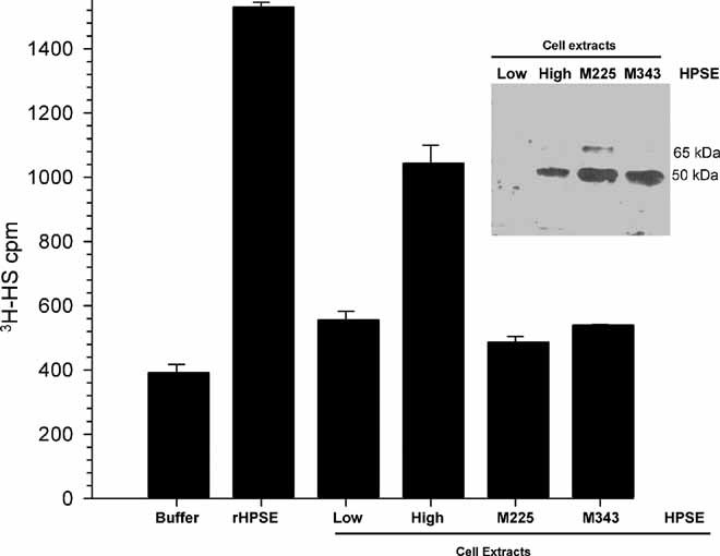
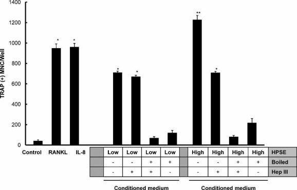
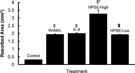
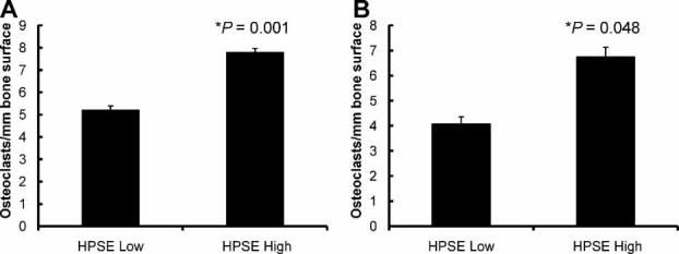
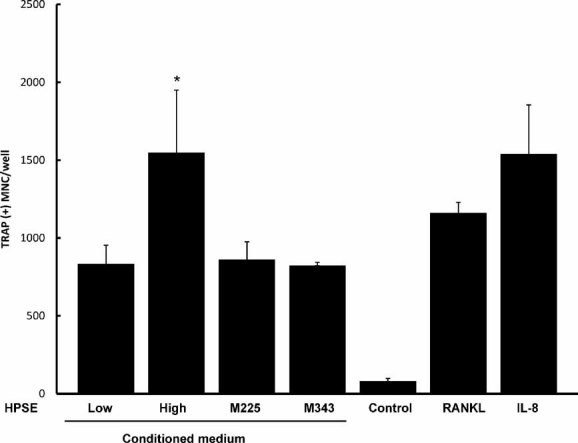
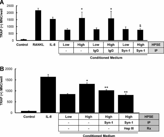
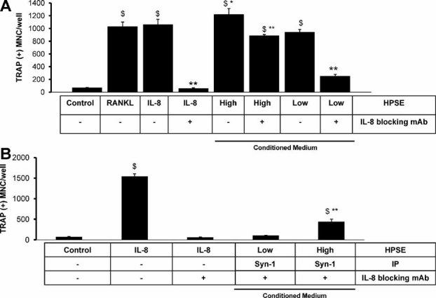
Tumor-stimulated bone resorption fuels tumor growth and marks a dramatic decline in the health and prognosis of breast cancer patients. Identifying mechanisms that mediate cross-talk between tumor and bone remains a key challenge. We previously demonstrated that breast cancer cells expressing high levels of heparanase exhibit enhanced shedding of the syndecan-1 proteoglycan. Moreover, when these heparanase-high cells are implanted in the mammary fat pad, they elevate bone resorption. In this study, conditioned medium from breast cancer cells expressing high levels of heparanase was shown to significantly stimulate human osteoclastogenesis in vitro (p < .05). The osteoclastogenic activity in the medium of heparanase-high cells was traced to the presence of syndecan-1, intact heparan sulfate chains, and heat-labile factor(s), including the chemokine interleukin 8 (IL-8). The enhanced osteoclastogenesis promoted by the heparanase-high cells results in a dramatic increase in bone resorption in vitro. In addition, the long bones of animals bearing heparanase-high tumors in the mammary fat pad had significantly higher numbers of osteoclasts compared with animals bearing tumors expressing low levels of heparanase (p < .05). Together these data suggest that syndecan-1 shed by tumor cells exerts biologic effects distal to the primary tumor and that it participates in driving osteoclastogenesis and the resulting bone destruction.


Figures

Fig. 1
Fig. 1
Heparanase protein levels and enzyme activity. The graph shows the heparan sulfate–degrading activity of the reaction buffer (Buffer), recombinant enzymatically active human heparanase (rHPSE, 1 µg), and cell extracts of HPSE-Low cells, HPSE-High cells, and cells expressing enzymatically inactive heparanase (M225 and M343). (Inset) Western blots of cell extracts probed with antibody to human heparanase. At the exposure shown, the most prominent band is the 50-kDa fully processed form of the enzyme in HPSE-High cells (High) and the two mutants (M225 and M343). Each lane was equivalently loaded for protein (30 µg).
Fig. 2
Fig. 2
Conditioned medium from HPSE-High cells stimulates osteoclastogenesis. An in vitro osteoclastogenesis assay was used to evaluate conditioned medium from HPSE-Low (left side) and HPSE-High cells (right side), as indicated below the bars (HPSE). Other treatments of the medium prior to osteoclastogenesis (HepIII or boiled) are also indicated below the bars. Medium from HPSE-Low cells, as well as treatment with RANKL or IL-8, produced significant osteoclastogenesis compared with csf-1-only control (p < .05 indicated by single asterisk). Medium from HPSE-High cells was significantly higher than that of HPSE-Low cells (p < .05 indicated by double asterisk). HepIII-treated conditioned medium from HPSE-High cells reduced osteoclast formation to the extent that it was not significantly different from the osteoclastogenic activity of the conditioned medium from the HPSE-Low cells but was still higher than csf-1 control. Boiling the conditioned medium or boiling after HepIII treatment completely abolished the osteoclastogenesis activity of the conditioned medium from either the HPSE-High or HPSE-Low cells down to csf-1 control levels. Results are indicative of at least three replicate experiments.
Fig. 3
Fig. 3
Medium from HPSE-High cells enhances bone resorption. Quantification of bone resorption on bone slices. Human osteoclasts were differentiated from PBMCs grown on bone and exposed to cell culture medium that was not conditioned by breast cancer cells (Control) or medium with addition of, RANKL (RANKL), IL-8 (IL-8), or medium conditioned by HPSE-High cells (HPSE-High), and medium conditioned by HPSE-Low cells (HPSE-Low). The $ indicates that all treatments are significantly different from csf-1 control (p < 0.05). The asterisk indicates that HPSE-High conditioned medium produced significantly more bone resorption than the all other conditions with p < .05. Results are indicative of at least two replicate experiments.
Fig. 4
Fig. 4
Orthotopic HPSE-High breast tumors increase osteoclast numbers in long bones. Femurs from animals bearing mammary fat pad tumors formed by HPSE-High or HPSE-Low cells were stained for TRACP, and the numbers of TRACP+ multinucleated cells were determined (A, experiment 1, n = 5 animals per group; B, experiment 2, n = 4 animals per group). A minimum of three sections of bone per animal were counted. Error bars represent standard error, and statistical significance was determined by a two-tailed t test. Significance was identified by p < .05.
Fig. 5
Fig. 5
Enzymatically active heparanase is required for stimulation of osteoclastogenesis. Media conditioned by cells expressing enzymatically active or inactive heparanase were tested for the capacity to stimulate osteoclastogenesis in vitro. Medium from HPSE-Low cells induced osteoclastogenesis that was significantly greater than csf-1 control (*p < .05) and not different from M225, and M343 cells expressing inactive heparanases. The osteoclastogenic activity of HPSE-High cells was significantly greater than the other heparanase-expressing cells ($, p < .05). As a positive control, osteoclastogenesis induced following the addition of RANKL or IL-8 is also significantly different from control (*p < .05). Results are indicative of at least three replicate experiments.
Fig. 6
Fig. 6
Shed syndecan-1 in conditioned medium of HPSE-High cells is required for enhanced osteoclastogenesis. (A) Osteoclastogenesis was increased significantly compared with control (csf-1 alone) in media conditioned by cells expressing low or high heparanase, as well as by the addition of IL-8 or RANKL (p < .05). Moreover, as expected, osteoclastogenesis was significantly higher for HPSE-high conditioned medium than for HPSE-low medium (*p < .05). However, only osteoclastogenesis stimulated by conditioned medium from cells expressing high levels of heparanase was decreased significantly by immunoprecipitation with a specific antibody to syndecan-1 (Syn-1, $) but not by immunoprecipitation with IgG. Results are indicative of at least three replicate experiments. (B) Immunoprecipitation of syndecan-1 from medium conditioned by heparanase-high cells significantly reduced the number of osteoclasts compared with medium without immunoprecipitation (**p < .05). Similalrly, immunoprecipitation of syndecan-1 from medium followed by HepIII treatment of medium also reduced the number of osteoclasts significantly compared with HPSE-High conditioned medium alone (**p < .05). HepIII treatment did not significantly reduce osteoclastogenesis compared with medium that lacked syndecan-1. Results are indicative of at least two replicate experiments.
Fig. 7
Fig. 7
IL-8 is a heparan sulfate–binding cytokine that cooperates with shed syndecan-1 to promote osteoclastogenesis. (A) Addition of IL-8 or RANKL significantly increases osteoclast formation compared with csf-1 control, as does medium from HPSE-High or -Low cells ($, p < .05). Treatment with a neutralizing IL-8 antibody significantly decreased osteoclast formation in all groups (**) except RANKL. Treatment of heparanase-low cells with IL-8 antibody was significantly different from all other conditioned media groups (**p < .05). Conditioned medium from HPSE-High cells treated with IL-8 antibody retained significantly higher osteoclastogenesis than csf-1 control ($, p < .05). Addition of the IL-8 antibody significantly reduced the osteoclastogenesis relative to untreated HPSE-High conditioned medium (**p < .05); therefore, this data point is marked ($**). (B) Addition of IL-8 significantly increased osteoclast formation compared with csf-1 control ($, p < .05). The increased osteoclastogenesis was completely abolished by addition of the IL-8 function-blocking monoclonal antibody. Blocking IL-8 and depleting syndecan-1 dramatically reduced the osteoclastogenesis of both HPSE-Low and HPSE-High conditioned medium. The osteoclastogenic activity of HPSE-High conditioned medium was decreased significantly by IL-8 blockade and syndecan-1 depletion relative the IL-8-positive control (**) but remained significantly different from csf-1 control ($**). Results are indicative of at least two replicate experiments.

Similar articles

Cited by

References

    1. Kakonen SM, Mundy GR. Mechanisms of osteolytic bone metastases in breast carcinoma. Cancer. 2003;97:834–839. - PubMed
    1. Guise TA, Yin JJ, Taylor SD, et al. Evidence for a causal role of parathyroid hormone-related protein in the pathogenesis of human breast cancer-mediated osteolysis. J Clin Invest. 1996;98:1544–1549. - PMC - PubMed
    1. Bendre MS, Gaddy-Kurten D, Mon-Foote T, et al. Expression of interleukin 8 and not parathyroid hormone-related protein by human breast cancer cells correlates with bone metastasis in vivo. Cancer Res. 2002;62:5571–5579. - PubMed
    1. Kang Y, Siegel PM, Shu W, et al. A multigenic program mediating breast cancer metastasis to bone. Cancer Cell. 2003;3:537–549. - PubMed
    1. Suva L, Griffin R, Makhoul I. Mechanisms of bone metastases of breast cancer. Endocr Relat Cancer. 2009;16:703–713. - PMC - PubMed

Publication types

MeSH terms