. 2010 Jun;25(6):1196-207.
doi: 10.1002/jbmr.29.

rBMP represses Wnt signaling and influences skeletal progenitor cell fate specification during bone repair

Affiliations

rBMP represses Wnt signaling and influences skeletal progenitor cell fate specification during bone repair

Steve Minear et al. J Bone Miner Res. 2010 Jun.

Abstract

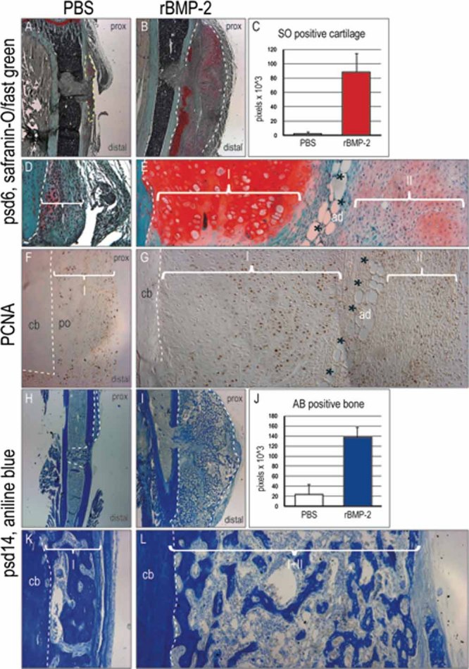
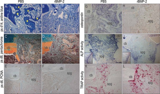
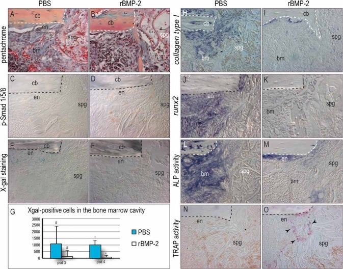
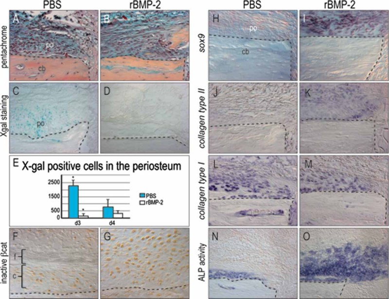
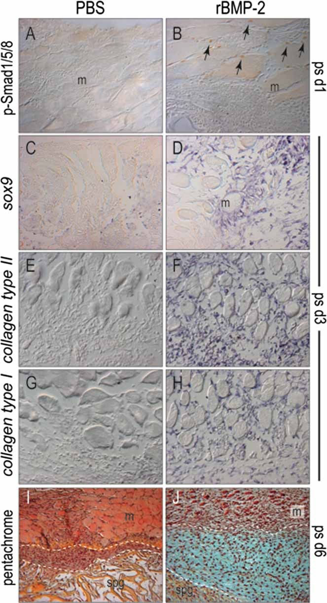
Bone morphogenetic proteins (BMPs) participate in multiple stages of the fetal skeletogenic program from promoting cell condensation to regulating chondrogenesis and bone formation through endochondral ossification. Here, we show that these pleiotropic functions are recapitulated when recombinant BMPs are used to augment skeletal tissue repair. In addition to their well-documented ability to stimulate chondrogenesis in a skeletal injury, we show that recombinant BMPs (rBMPs) simultaneously suppress the differentiation of skeletal progenitor cells in the endosteum and bone marrow cavity to an osteoblast lineage. Both the prochondrogenic and antiosteogenic effects are achieved because rBMP inhibits endogenous beta-catenin-dependent Wnt signaling. In the injured periosteum, this repression of Wnt activity results in sox9 upregulation; consequently, cells in the injured periosteum adopt a chondrogenic fate. In the injured endosteum, rBMP also inhibits Wnt signaling, which results in the runx2 and collagen type I downregulation; consequently, cells in this region fail to differentiate into osteoblasts. In muscle surrounding the skeletal injury site, rBMP treatment induces Smad phosphorylation followed by exuberant cell proliferation, an increase in alkaline phosphatase activity, and chondrogenic differentiation. Thus different populations of adult skeletal progenitor cells interpret the same rBMP stimulus in unique ways, and these responses mirror the pleiotropic effects of BMPs during fetal skeletogenesis. These mechanistic insights may be particularly useful for optimizing the reparative potential of rBMPs while simultaneously minimizing their adverse outcomes.


Figures

Fig. 1
Fig. 1
rBMP-2 induces a large extracortical callus via endochondral ossification. (A) safranin-O/fast green staining on postoperative day 6 cartilage in the PBS-treated injured periosteum signaling endochondral ossification (dotted lines). (B) rBMP-2 treatment induces a large cartilage callus superficial to the cortex. (C) Histomorphometric measurements show that compared with PBS, rBMP-2 induces a 31-fold increase in the size of the cartilage callus. (D) A ×40 view of safranin-O/fast green staining reveals minimal cartilage in the periosteal reaction (po) to injury in PBS-treated samples. (E) rBMP-2 treatment induces two large cartilaginous domains. One is the periosteal reaction (I) and the other is superficial (II) and is separated from one another by adipose/fibrous tissue (asterisks). (F) PCNA staining reveals proliferation in both the supraperiosteum and periosteum in PBS-treated controls. (G) rBMP-2 treatment reveals two domains of proliferating cells, a periosteal reaction and a second, supraperiosteal response. (H) Postoperative day 14 aniline blue staining of control samples shows robust osteogenesis in the bone marrow cavity (dotted line). (I) rBMP-2-treated samples exhibit a large bony callus located exclusively in the extracortical space; bone formation in the injury site is not detectable. (J) rBMP-2 treatment induces a 6-fold increase in the size of the bony callus. (K) In PBS-treated samples, aniline blue staining reveals the periosteal reaction on postoperative day 14. (L) By postoperative day 14, the two cartilage domains have coalesced into a single bony domain in the rBMP-2-treated samples (white bracket). ps = postsurgical; ad = adipogenic; cb = cortical bone; is = injury site.
Fig. 2
Fig. 2
rBMP-2 does not induce bone formation in the marrow cavity. (A) In controls, new bone forms primarily in the bone marrow cavity, which begins to bridge the defect (asterisks), but (B) in rBMP-2-treated samples, there is no evidence of osteoid matrix in this location. (C, D) Pentachrome staining on postoperative day 6 reveals no obvious differences in the placement of the collagen sponge, the extent of cellular infiltrate, or the amount of vascularization between control and rBMP-2-treated samples. Despite their histologic equivalency, (E, F) the number of PCNA-immunopositive cells is increased in controls compared with rBMP-2-treated samples. In addition, (G, H) controls show higher levels of osteopontin expression than rBMP-2-treated samples. (I) PBS-treated controls also show more extensive alkaline phosphatase activity than (J) rBMP-2-treated samples. Insets in panels I and J illustrate equivalent levels of alkaline phosphatase staining in the growth plates of both tissue sections. In contrast, (K, L) TRAP activity is reduced in controls compared with rBMP-2-treated samples. Dotted line outlines cortical bone. ps = postsurgical; cb = cortical bone; spg = sponge.
Fig. 3
Fig. 3
rBMP-2 suppresses differentiation of osteoprogenitors in the bone marrow cavity. (A, B) Pentachrome staining of the bone marrow cavity on postoperative day 3 shows evidence of the collagen sponge carrier as well as a dense cellular mass with abundant red blood cells. PBS-treated controls show more extracellular matrix than rBMP-2-treated samples. (C) Immunostaining for phospho-Smad 1/5/8 reveals no cells responding to a BMP-2 stimulus. (D) More cells, relative to controls, are responding to rBMP-2 in the endosteum of rBMP-2-treated samples on postoperative day 3. (E) Axin2lacZ/+ mice were used to map β-catenin-dependent Wnt signaling in the bone marrow cavity. PBS-treated control endosteum demonstrates extensive Wnt responsiveness. (F) rBMP-2 abrogates the β-catenin-dependent Wnt responsiveness in the endosteum. (G) Quantification reveals a statistically significant reduction of Wnt responsiveness on postoperative days 3 and 4. (H, J) Control samples demonstrate expression of early markers of osteogenesis, including collagen type I and runx2, whereas (I, K) rBMP-2-treated samples exhibit lower levels of gene expression. (L) PBS-treated samples show robust alkaline phosphatase activity in the marrow space, but (M) rBMP-2 downregulates this activity. (N, O) rBMP-2-treated samples exhibit more TRAP activity than PBS-treated samples. Dotted line outlines cortical bone. cb = cortical bone; en = endosteum; spg = sponge; bm = bone marrow. #p < .05; *p < .01.
Fig. 4
Fig. 4
rBMP-2 induces differentiation of chondrocytes and upregulates osteochondroprogenitor markers in the extracortical space. On postoperative day 3, (A, B) pentachrome staining reveals a similar periosteal reaction in PBS- and rBMP-2-treated samples. (C) Axin2lacZ/+ mice show an extensive distribution of β-catenin Wnt responsiveness in the injured periosteum. (D) rBMP-2 treatment abrogates this Wnt responsiveness here. (E) Quantification reveals a statistically significant reduction of Xgal+ cells on postoperative day 3. This difference is no longer present by day 4. (F) PBS-treated periosteum demonstrates minimal sox9 expression of day 3. (G) rBMP-2-treated samples demonstrate a more robust and wider distribution of sox9 expression in the periosteum. (H, I) Relative to PBS treatment, rBMP-2-treated samples exhibit higher collagen type II expression in the periosteum. (J, K) PBS- and rBMP-2-treated samples show similar collagen type I expression in the periosteum. (L, M) Relative to PBS-treated controls, rBMP-2-treated samples exhibit higher alkaline phosphatase activity in the periosteum. po = periosteum. *p < .01.
Fig. 5
Fig. 5
rBMP-2 induces osteochondroprogenitor markers in the supraperiosteal/muscle compartment. (A) PBS-treated muscle does not exhibit any Smad 1/5/8 phosphorylation on postoperative day 1. (B) rBMP-2 treatment induces phospho-Smad 1/5/8 immunostaining in this tissue by day 1 (arrowheads). (C, D) On postoperative day 3, pentachrome staining of both PBS- and rBMP-2-treated supraperiosteum reveals a highly cellular region with no overt osteogenesis or chondrogenesis in the supraperiosteal space. (E) PBS samples do not express collagen type I. (F) rBMP-2 treatment induces collagen type I expression. (G) PBS-treated samples do not express collagen type II. (H) rBMP-2 induces collagen type II expression here. (I) When the injury is modified to exclude the bone marrow cavity and separate the periosteal/supraperiosteal compartments, control samples exhibit only fibrous tissue between sponge and muscle (outlined). (J) rBMP-2-treated samples induce a separate chondrogenic response from supraperiosteal tissue in the defects. m = muscle; spg = sponge.
Fig. 6
Fig. 6
Tissue-specific responses to rBMP-2 can be used to predict the relative success of in vivo applications for the growth factor. (A) Following implantation, three populations of cells respond to rBMP: (1) cells in the bone marrow cavity, (2) cells in the injured periosteum, and (3) cells in the muscle overlying the injury site. Cells in the periosteum and endosteum are typically Wnt responsive (blue cells); rBMP treatment abrogates this responsiveness (red cells). (B) At an intermediate time point (in a mouse model, between 6 and 10 days after surgery), rBMP elicits three separate responses: (1) cells in the bone marrow subsequent to reduced Wnt signaling, neither proliferate nor differentiate into osteoblasts, (2) osteochondroprogenitor cells in the injured periosteum, which also exhibited a reduction in Wnt responsiveness at early time points, respond to rBMP by expressing sox9 and adopting a chondrogenic fate, and (3) cells in the muscle respond to rBMP by adopting a chondrogenic fate, which contributes to the callus size. (C) At later stages of repair (in mice, around day 14), (1) the collagen sponge has resorbed, yet there is still no evidence of an osteogenic response from bone marrow cells, and (2) a coalescence of the periosteal and supraperiosteal cells creates a large extracortical bony bridge via the process of endochondral ossification.

Comment in

Similar articles

Cited by

References

    1. Akiyama H, Chaboissier MC, Martin JF, Schedl A, de Crombrugghe B. The transcription factor Sox9 has essential roles in successive steps of the chondrocyte differentiation pathway and is required for expression of Sox5 and Sox6. Genes Dev. 2002;16:2813–2828. - PMC - PubMed
    1. Ducy P, Zhang R, Geoffroy V, Ridall AL, Karsenty G. Osf2/Cbfa1: a transcriptional activator of osteoblast differentiation. Cell. 1997;89:747–754. - PubMed
    1. Komori T, Yagi H, Nomura S, et al. Targeted disruption of Cbfa1 results in a complete lack of bone formation owing to maturational arrest of osteoblasts. Cell. 1997;89:755–764. - PubMed
    1. Kobayashi T, Lu J, Cobb BS, et al. Dicer-dependent pathways regulate chondrocyte proliferation and differentiation. Proc Natl Acad Sci U S A. 2008;105:1949–1954. - PMC - PubMed
    1. Vortkamp A, Lee K, Lanske B, Segre GV, Kronenberg HM, Tabin CJ. Regulation of rate of cartilage differentiation by Indian hedgehog and PTH-related protein. Science. 1996;273:613–622. - PubMed

Publication types

MeSH terms