

Mechanical implications of estrogen supplementation in early postmenopausal women
- PMID: 20200948
- PMCID: PMC3153138
- DOI: 10.1002/jbmr.33
Mechanical implications of estrogen supplementation in early postmenopausal women
Abstract
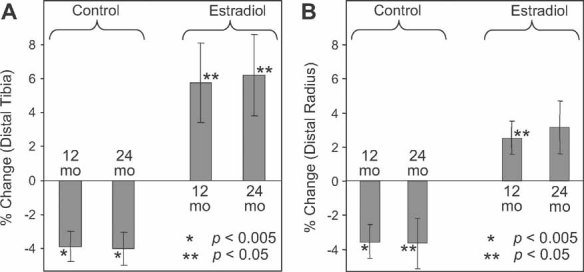
Whereas the structural implications of drug intervention are well established, there are few data on the possible mechanical consequences of treatment. In this work we examined the changes in elastic and shear moduli (EM and SM) in a region of trabecular bone in the distal radius and distal tibia of early postmenopausal women on the basis of MRI-based micro-finite-element (microFE) analysis. Whole-section axial stiffness (AS) encompassing both trabecular and cortical compartments was evaluated as well. The study was conducted on previously acquired high-resolution images at the two anatomic sites. Images were processed to yield a 3D voxel array of bone-volume fraction (BVF), which was converted to a microFE model of hexahedral elements in which tissue modulus was set proportional to voxel BVF. The study comprised 65 early postmenopausal women (age range 45 to 55 years), of whom 32 had chosen estrogen supplementation (estradiol group); the remainder had not (control group). Subjects had been scanned at baseline and 12 and 24 months thereafter. At the distal tibia, EM and SM were reduced by 2.9% to 5.5% in the control group (p < .05 to <.005), but there was no change in the estradiol subjects. AS decreased 3.9% (4.0%) in controls (p < .005) and increased by 5.8% (6.2%) in estradiol group subjects (p < .05) at 12 (24) months. At the distal radius, EM and SM changes from baseline were not significant, but at both time points AS was increased in estradiol group subjects and decreased in controls (p < .005 to <.05), albeit by a smaller margin than at the tibia. EM and SM were strongly correlated with BV/TV (r(2) = 0.44 to 0.92) as well as with topologic parameters expressing the ratio of plates to rods (r(2) = 0.45 to 0.82), jointly explaining up to 96% of the variation in the mechanical parameters. Finally, baseline AS was strongly correlated between the two anatomic sites (r(2) = 0.58), suggesting that intersubject variations in the bone's mechanical competence follows similar mechanisms. In conclusion, the results demonstrate that micro-MRI-based microFE models are suited for the study of the mechanical implications of antiresorptive treatment. The data further highlight the anabolic effect of short-term estrogen supplementation.
(c) 2010 American Society for Bone and Mineral Research.
Figures


Similar articles
-
In vivo assessment of architecture and micro-finite element analysis derived indices of mechanical properties of trabecular bone in the radius.Osteoporos Int. 2002 Jan;13(1):6-17. doi: 10.1007/s198-002-8332-0. Osteoporos Int. 2002. PMID: 11878456
-
In vivo magnetic resonance detects rapid remodeling changes in the topology of the trabecular bone network after menopause and the protective effect of estradiol.J Bone Miner Res. 2008 May;23(5):730-40. doi: 10.1359/jbmr.080108. J Bone Miner Res. 2008. PMID: 18251704 Free PMC article.
-
Accuracy of high-resolution in vivo micro magnetic resonance imaging for measurements of microstructural and mechanical properties of human distal tibial bone.J Bone Miner Res. 2010 Sep;25(9):2039-50. doi: 10.1002/jbmr.92. J Bone Miner Res. 2010. PMID: 20499379 Free PMC article.
-
Computational biomechanics of the distal tibia from high-resolution MR and micro-CT images.Bone. 2010 Sep;47(3):556-63. doi: 10.1016/j.bone.2010.05.039. Epub 2010 May 31. Bone. 2010. PMID: 20685323 Free PMC article.
-
In vivo estimation of bone stiffness at the distal femur and proximal tibia using ultra-high-field 7-Tesla magnetic resonance imaging and micro-finite element analysis.J Bone Miner Metab. 2012 Mar;30(2):243-51. doi: 10.1007/s00774-011-0333-1. Epub 2011 Nov 30. J Bone Miner Metab. 2012. PMID: 22124539 Free PMC article.
Cited by
-
Computationally-optimized bone mechanical modeling from high-resolution structural images.PLoS One. 2012;7(4):e35525. doi: 10.1371/journal.pone.0035525. Epub 2012 Apr 25. PLoS One. 2012. PMID: 22558164 Free PMC article.
-
Finite element analysis applied to 3-T MR imaging of proximal femur microarchitecture: lower bone strength in patients with fragility fractures compared with control subjects.Radiology. 2014 Aug;272(2):464-74. doi: 10.1148/radiol.14131926. Epub 2014 Apr 2. Radiology. 2014. PMID: 24689884 Free PMC article.
-
Potential of in vivo MRI-based nonlinear finite-element analysis for the assessment of trabecular bone post-yield properties.Med Phys. 2013 May;40(5):052303. doi: 10.1118/1.4802085. Med Phys. 2013. PMID: 23635290 Free PMC article.
-
Advances in imaging approaches to fracture risk evaluation.Transl Res. 2017 Mar;181:1-14. doi: 10.1016/j.trsl.2016.09.006. Epub 2016 Oct 17. Transl Res. 2017. PMID: 27816505 Free PMC article. Review.
-
Quantitative assessment of trabecular bone micro-architecture of the wrist via 7 Tesla MRI: preliminary results.MAGMA. 2011 Aug;24(4):191-9. doi: 10.1007/s10334-011-0252-0. Epub 2011 May 5. MAGMA. 2011. PMID: 21544680 Free PMC article.
References
-
- Seeman E, Delmas PD. Bone quality–the material and structural basis of bone strength and fragility. N Engl J Med. 2006;354:2250–61. - PubMed
-
- Paschalis EP, Boskey AL, Kassem M, Eriksen EF. Effect of hormone replacement therapy on bone quality in early postmenopausal women. J Bone Miner Res. 2003;18:955–9. - PubMed
-
- Recker RR, Barger-Lux MJ. The elusive concept of bone quality. Curr Osteoporos Rep. 2004;2:97–100. - PubMed
-
- Sarkar S, Mitlak BH, Wong M, Stock JL, Black DM, Harper KD. Relationships between bone mineral density and incident vertebral fracture risk with raloxifene therapy. J Bone Miner Res. 2002;17:1–10. - PubMed
-
- Genant HK, Jiang Y. Advanced imaging assessment of bone quality. Ann N Y Acad Sci. 2006;1068:410–28. - PubMed
Publication types
MeSH terms
Substances
Grants and funding
LinkOut - more resources
Full Text Sources
Medical
Research Materials
