. 2010 Jul;25(7):1668-79.
doi: 10.1002/jbmr.37.

Cell-based immunotherapy with mesenchymal stem cells cures bisphosphonate-related osteonecrosis of the jaw-like disease in mice

Affiliations

Cell-based immunotherapy with mesenchymal stem cells cures bisphosphonate-related osteonecrosis of the jaw-like disease in mice

Takashi Kikuiri et al. J Bone Miner Res. 2010 Jul.

Abstract

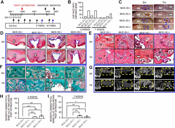
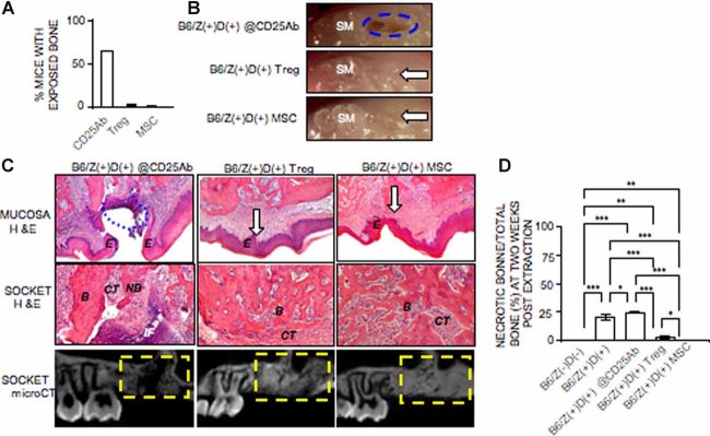
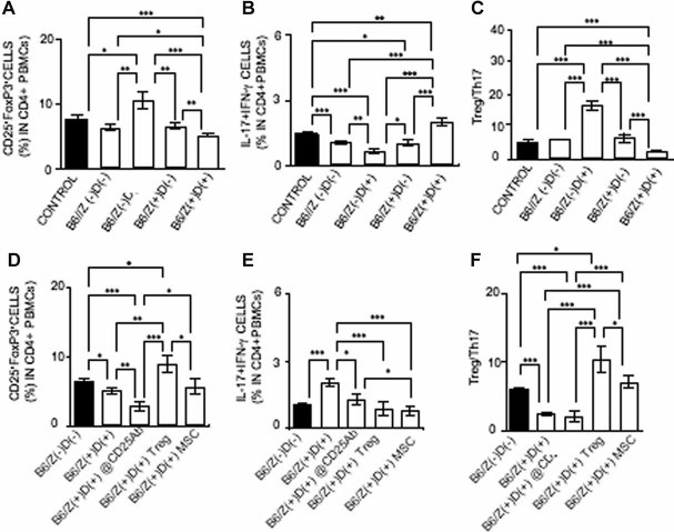
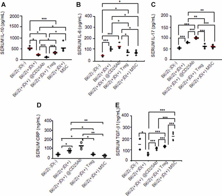
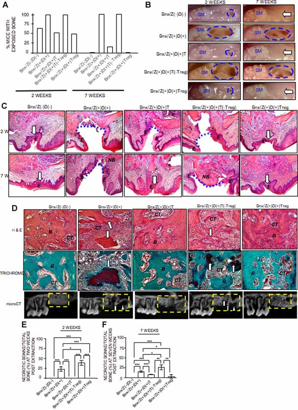
Patients on high-dose bisphosphonate and immunosuppressive therapy have an increased risk of bisphosphonate-related osteonecrosis of the jaw (BRONJ); despite the disease severity, its pathophysiology remains unknown, and appropriate therapy is not established. Here we have developed a mouse model of BRONJ-like disease that recapitulates major clinical and radiographic manifestations of the human disease, including characteristic features of an open alveolar socket, exposed necrotic bone or sequestra, increased inflammatory infiltrates, osseous sclerosis, and radiopaque alveolar bone. We show that administration of zoledronate, a potent aminobisphosphonate, and dexamethasone, an immunosuppressant drug, causes BRONJ-like disease in mice in part by suppressing the adaptive regulatory T cells, Tregs, and activating the inflammatory T-helper-producing interleukin 17 cells, Th17. Most interestingly, we demonstrate that systemic infusion with mesenchymal stem cells (MSCs) prevents and cures BRONJ-like disease possibly via induction of peripheral tolerance, shown as an inhibition of Th17 and increase in Treg cells. The suppressed Tregs/Th17 ratio in zoledronate- and dexamethasone-treated mice is restored in mice undergoing salvage therapy with Tregs. These findings provide evidence of an immunity-based mechanism of BRONJ-like disease and support the rationale for in vivo immunomodulatory therapy using Tregs or MSCs to treat BRONJ.


Figures

Fig. 1
Fig. 1
Development of BRONJ-like lesions in C57BL/6J mice. (A) Experimental protocol of inducing a BRONJ-like murine model. C57BL/6J (B6) mice were intravenously injected with Zol (125 µg/kg) and Dex (5 mg/kg) twice weekly for 1 week before surgical extraction of the left maxillary first molar and continuous injection of Zol and Dex twice weekly for 2 or 7 weeks. (B) Incidence of BRONJ-like lesion, manifested as an unhealed open socket with an area of exposed bone and no mucosal coverage, at the extraction site in B6 mice treated with Zol (Z) and Dex (D) (B6/Z+D+) for 3 and 8 weeks, whereby mice with no treatment (B6/ZD), Dex only (B6/ZD+), and Zol only (B6/Z+D) served as controls. (C) Representative gross clinical appearance of gingival mucosa at the extraction sites at 2 and 7 weeks after tooth extraction. Blue circles represent images of the apparent mucosal disruption with exposed bone at the extracted site adjacent to the second molar (SM), whereas open arrows point to the healed gingival mucosa. (D) Histologic images of extraction sockets showing open sockets without epithelial lining (blue dot line) and healed gingival mucosa with complete epithelial coverage (open arrow). NB = necrotic bone. (E, F) H&E (E) and trichrome (F) staining of extraction socket areas displaying newly formed bone (B), connective tissues (CT), necrotic bone (NB), and inflammatory cell infiltrates (IF); open arrows point to the necrotic bones. (G) µCT analysis showing necrotic bone (open arrow) and reduced bone formation (triangle) in B6/Z+D+ mice. (H, I) Quantification of necrotic bone areas in B6/Z+D+ and B6/ZD groups at 2 and 7 weeks after tooth extraction. *p < .05; ***p < .005. The results are representative of three independent experiments and are shown as mean ± SD.
Fig. 2
Fig. 2
Treg and MSC treatment prevents development of BRONJ-like lesions and enhances new bone formation in the C57BL/6J mouse model. (A) Incidence of BRONJ-like lesions, manifested as an unhealed open socket with area of exposed bone and no mucosal coverage, at the extraction site in Zol/Dex-treated C57BL/6J mice receiving ip injections of anti-CD25 antibody (CD25Ab) (B6/Z+D+@CD25Ab) or systemic infusion of Tregs (B6/Z+D+Treg) or MSCs (B6/Z+D+MSC) at 7 weeks after tooth extraction. CD25Ab was administered 2 days before tooth extraction. Treg and MSC infusions were initiated 2 days after tooth extraction. (B) Representative gross clinical appearance of gingival mucosa at the extraction sites at 7 weeks after tooth extraction. Blue circles represent images of the apparent mucosal disruption, with exposed bone at the extracted site adjacent to the second molar (SM), whereas open arrows point to the healed gingival mucosa. (C) Histologic analysis showing an open socket without epithelial lining (blue dot line), healed gingival mucosa with complete epithelial coverage (open arrow) at the extraction sites (upper panel), and newly formed bone (B), connective tissues (CT), necrotic bone (NB), and inflammatory cell infiltrates (IF) (middle panels); open arrows point to the necrotic bones; µCT analysis (lower panel) showing necrotic bone and reduced bone formation in Zol/Dex-treated C57BL/6J mice receiving ip injections of CD25 antibody (B6/Z+D+@CD25Ab) but not in mice treated with systemic infusion of Tregs (B6/Z+D+Treg) or MSCs (B6/Z+D+MSC). (D) Quantification of necrotic bone areas in Zol/Dex-treated C57BL/6J mice receiving ip injection of CD25Ab or systemic infusion of Tregs and MSCs at 2 weeks after tooth extraction. *p < .05; **p < .01; ***p < .005. The results are representative of three independent experiments.
Fig. 3
Fig. 3
Effects of various treatments on T cell profile of CD4+CD25+Foxp3+ Tregs and CD4+IL17+IFNγ Th17 cells in BRONJ-like C57BL/6J mice. (A, D) Profile of CD4+CD25+Foxp3+ Tregs in peripheral blood of C57BL/6J mice treated with Zol (Z) and Dex (D) (B6/Z+D+) (A), Zol/Dex and ip injection of CD25Ab (B6/Z+D+@CD25Ab), Zol/Dex and systemic infusion of Tregs (B6/Z+D+Treg), or MSCs (B6/Z+D+MSC) (D) at 2 weeks after tooth extraction; mice with or without tooth extraction (control) and no drug treatment (B6/ZD), Dex only (B6/ZD+), and Zol only (B6/Z+D) served as controls. *p < .05; **p < .01; ***p < .001. (B, E) Profile of CD4+IL17+IFNγ cells in peripheral blood of C57BL/6J mice treated with Zol (Z) and Dex (D) (B6/Z+D+) (B), Zol/Dex and ip injection of CD25Ab (B6/Z+D+@CD25Ab), Zol/Dex and systemic infusion of Tregs (B6/Z+D+Treg) or MSCs (B6/Z+D+MSC) (E) at 2 weeks after tooth extraction. Mice with no tooth extraction (control) or with tooth extraction but with no treatment (B6/ZD) served as controls. *p < .05; **p < .01; ***p < .005. (C, F) The ratio of CD4+CD25+Foxp3+ cells (Tregs) and CD4+IL17+IFNγ cells (Th17 cells) in peripheral blood of C57BL/6J mice after receiving different treatment regimens. *p < .05; ***p < .005. The results are representative of three independent experiments.
Fig. 4
Fig. 4
Effects of various treatments on serum inflammatory cytokine level in BRONJ-like C57BL/6J mice. C57BL/6J mice were treated with Zol (Z) and Dex (D) (B6/Z+D+), Zol/Dex and ip injection of CD25Ab (B6/Z+D+@CD25Ab), Zol/Dex and systemic infusion of Tregs (B6/Z+D+Treg), or MSCs (B6/Z+D+MSC) at 2 weeks after tooth extraction. Peripheral blood samples were collected, and serum levels of IL-10 (A), IL-6 (B), IL-17 (C), CRP (D), and total TGF-β1 (E) were determined by ELISA. Mice with no tooth extraction or with tooth extraction but with no treatment (B6/ZD) served as controls. *p < .05; **p < .01; ***p < .005. The results are representative of three independent experiments.
Fig. 5
Fig. 5
Development of BRONJ-like lesions in immunocompromised mice. (A) Incidence of BRONJ-like lesion, manifested as an unhealed open socket with an area of exposed bone and no mucosal coverage, at the extraction site in Beige nu/nu Xid (III) (Bnx) mice treated with Zol (Z) alone (Bnx/Z+D) or in combination with Dex (D) (Bnx/Z+D+) for 3 and 8 weeks, whereby mice with no treatment (Bnx/ZD) served as controls. To test adaptive transfer of pan-T and Treg cells in the treatment of BRONJ-like lesion, three experimental groups were included: Bnx/Z+D+ with intravenous infusion of either pan-T cells (Bnx/Z+D+T) or T cells with depleted Tregs [Bnx/Z+D+T (–Treg)] or purified Tregs (Bnx/Z+D+Treg). T cell infusion was performed 2 days after tooth extraction. (B) Representative gross clinical appearance of gingival mucosa at the extraction sites at 2 and 7 weeks after tooth extraction. Blue circles represent images of the apparent mucosal disruption with exposed bone at the extracted site adjacent to the second molar (SM), whereas open arrows point to the healed gingival mucosa. (C) Histologic images of extraction sockets showing open socket without epithelial lining (blue dot line) and healed gingival mucosa with complete epithelial coverage (open arrow). NB = necrotic bone. (D) H&E (upper panel) and trichrome (middle panel) staining of extraction socket areas displaying newly formed bone (B), connective tissues (CT), and necrotic bone (NB) at 7 weeks after tooth extraction. Open arrows point to the necrotic bones. µCT analysis (lower panel) showed necrotic bone (open arrow) and reduced bone formation (triangle) in Bnx/Z+D+ and Bnx/Z+D+T (–Treg) groups. (E, F) Quantification of necrotic bone areas in Bnx/Z+D+ and mice treated with systemic infusion of different T cells at 2 and 7 weeks after tooth extraction. *p < .05; **p < .01; ***p < .005. Bars indicate SD. The results are representative of three independent experiments.

Similar articles

Cited by

References

    1. Lipton A. New therapeutic agents for the treatment of bone diseases. Expert Opin Biol Ther. 2006;5:817–832. - PubMed
    1. Berenson JR, Hillner BE, Kyle RA, et al. American Society of Clinical Oncology clinical practice guidelines: the role of bisphosphonates in multiple myeloma. J Clin Oncol. 2002;20:3719–3736. - PubMed
    1. Lipton A, Theriault RL, Hortobagyi GN, et al. Pamidronate prevents skeletal complications and is effective palliative treatment in women with breast carcinoma and osteolytic bone metastases: long-term follow-up of two randomized, placebo-controlled trials. Cancer. 2000;88:1082–1090. - PubMed
    1. Berenson JR, Rosen LS, Howell A, et al. Zoledronic acid reduces skeletal-related events in patients with osteolytic metastases. Cancer. 2001;91:1191–1200. - PubMed
    1. Marx RE. Pamidronate (Aredia) and zoledronate (Zometa) induced avascular necrosis of the jaws: a growing epidemic. J Oral Maxillofac Surg. 2003;61:1115–1117. - PubMed

Publication types

MeSH terms