

Thyroid hormone-mediated growth and differentiation of growth plate chondrocytes involves IGF-1 modulation of beta-catenin signaling
- PMID: 20200966
- PMCID: PMC3123724
- DOI: 10.1002/jbmr.5
Thyroid hormone-mediated growth and differentiation of growth plate chondrocytes involves IGF-1 modulation of beta-catenin signaling
Abstract
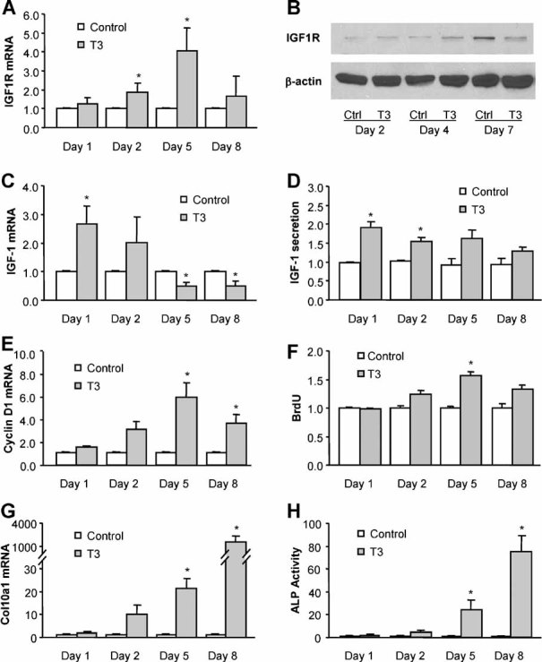
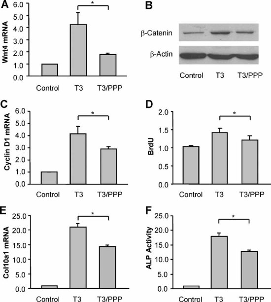
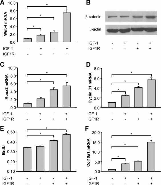
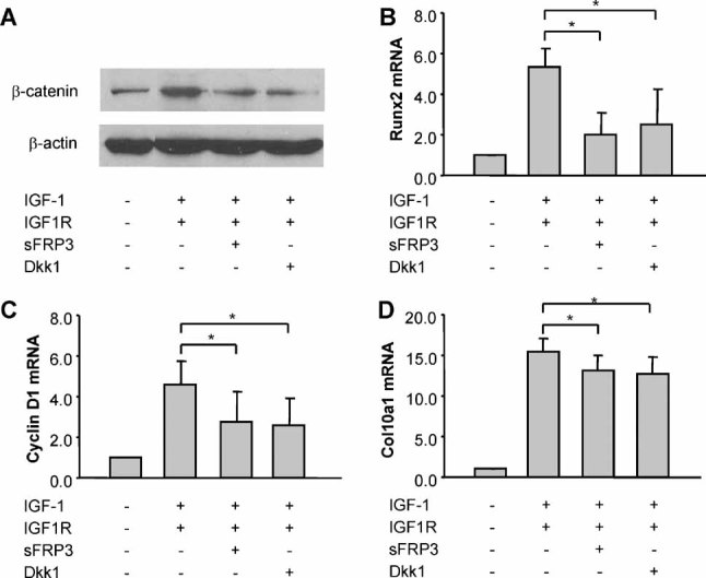
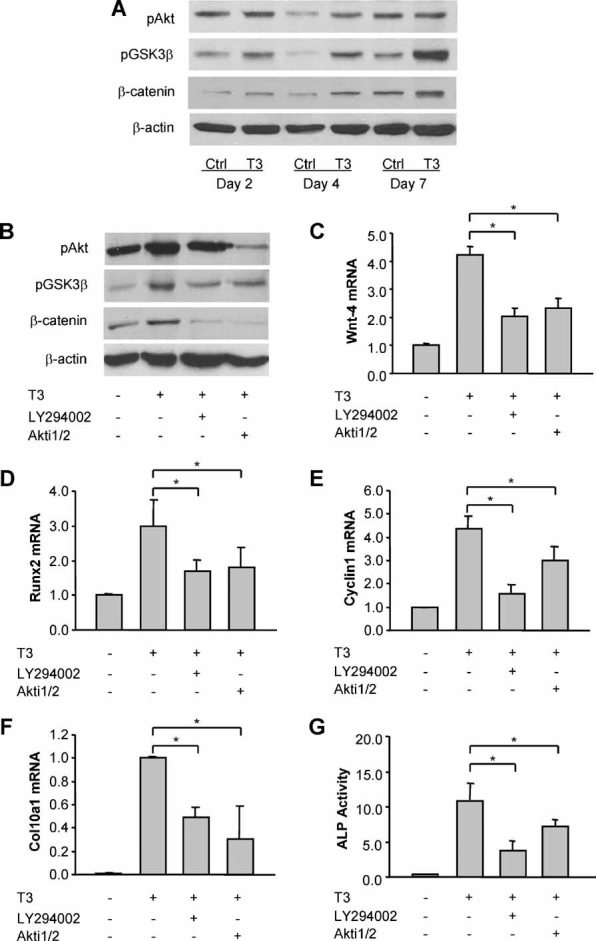
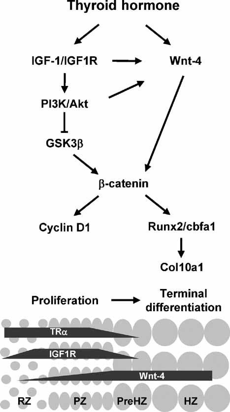
Thyroid hormone regulates terminal differentiation of growth plate chondrocytes in part through modulation of the Wnt/beta-catenin signaling pathway. Insulin-like growth factor 1 (IGF-1) has been described as a stabilizer of beta-catenin, and thyroid hormone is a known stimulator of IGF-1 receptor expression. The purpose of this study was to test the hypothesis that IGF-1 signaling is involved in the interaction between the thyroid hormone and the Wnt/beta-catenin signaling pathways in regulating growth plate chondrocyte proliferation and differentiation. The results show that IGF-1 and the IGF- receptor (IGF1R) stimulate Wnt-4 expression and beta-catenin activation in growth plate chondrocytes. The positive effects of IGF-1/IGF1R on chondrocyte proliferation and terminal differentiation are partially inhibited by the Wnt antagonists sFRP3 and Dkk1. T(3) activates IGF-1/IGF1R signaling and IGF-1-dependent PI3K/Akt/GSK-3beta signaling in growth plate chondrocytes undergoing proliferation and differentiation to prehypertrophy. T(3)-mediated Wnt-4 expression, beta-catenin activation, cell proliferation, and terminal differentiation of growth plate chondrocytes are partially prevented by the IGF1R inhibitor picropodophyllin as well as by the PI3K/Akt signaling inhibitors LY294002 and Akti1/2. These data indicate that the interactions between thyroid hormone and beta-catenin signaling in regulating growth plate chondrocyte proliferation and terminal differentiation are modulated by IGF-1/IGF1R signaling through both the Wnt and PI3K/Akt signaling pathways. While chondrocyte proliferation may be triggered by the IGF-1/IGF1R-mediated PI3K/Akt/GSK3beta pathway, cell hypertrophy is likely due to activation of Wnt/beta-catenin signaling, which is at least in part initiated by IGF-1 signaling or the IGF-1-activated PI3K/Akt signaling pathway.
(c) 2010 American Society for Bone and Mineral Research.
Figures
Similar articles
-
Thyroid hormone interacts with the Wnt/beta-catenin signaling pathway in the terminal differentiation of growth plate chondrocytes.J Bone Miner Res. 2007 Dec;22(12):1988-95. doi: 10.1359/jbmr.070806. J Bone Miner Res. 2007. PMID: 17708712
-
Leptin synergizes with thyroid hormone signaling in promoting growth plate chondrocyte proliferation and terminal differentiation in vitro.Bone. 2011 May 1;48(5):1022-7. doi: 10.1016/j.bone.2011.02.012. Epub 2011 Feb 22. Bone. 2011. PMID: 21349356
-
Carboxypeptidase Z (CPZ) links thyroid hormone and Wnt signaling pathways in growth plate chondrocytes.J Bone Miner Res. 2009 Feb;24(2):265-73. doi: 10.1359/jbmr.081014. J Bone Miner Res. 2009. PMID: 18847325 Free PMC article.
-
Are Wnt/β-Catenin and PI3K/AKT/mTORC1 Distinct Pathways in Colorectal Cancer?Cell Mol Gastroenterol Hepatol. 2020;10(3):491-506. doi: 10.1016/j.jcmgh.2020.04.007. Epub 2020 Apr 22. Cell Mol Gastroenterol Hepatol. 2020. PMID: 32334125 Free PMC article. Review.
-
Biology and pathology of Rho GTPase, PI-3 kinase-Akt, and MAP kinase signaling pathways in chondrocytes.J Cell Biochem. 2010 Jun 1;110(3):573-80. doi: 10.1002/jcb.22604. J Cell Biochem. 2010. PMID: 20512918 Free PMC article. Review.
Cited by
-
Relating the chondrocyte gene network to growth plate morphology: from genes to phenotype.PLoS One. 2012;7(4):e34729. doi: 10.1371/journal.pone.0034729. Epub 2012 Apr 30. PLoS One. 2012. PMID: 22558096 Free PMC article.
-
Altered IGF-I activity and accelerated bone elongation in growth plates precede excess weight gain in a mouse model of juvenile obesity.J Appl Physiol (1985). 2022 Feb 1;132(2):511-526. doi: 10.1152/japplphysiol.00431.2021. Epub 2022 Jan 6. J Appl Physiol (1985). 2022. PMID: 34989650 Free PMC article.
-
Hindlimb heating increases vascular access of large molecules to murine tibial growth plates measured by in vivo multiphoton imaging.J Appl Physiol (1985). 2014 Feb 15;116(4):425-38. doi: 10.1152/japplphysiol.01212.2013. Epub 2013 Dec 26. J Appl Physiol (1985). 2014. PMID: 24371019 Free PMC article.
-
The Regulatory Role of Signaling Crosstalk in Hypertrophy of MSCs and Human Articular Chondrocytes.Int J Mol Sci. 2015 Aug 14;16(8):19225-47. doi: 10.3390/ijms160819225. Int J Mol Sci. 2015. PMID: 26287176 Free PMC article. Review.
-
Thyroid Hormones in Early Pregnancy and Birth Weight: A Retrospective Study.Biomedicines. 2025 Feb 21;13(3):542. doi: 10.3390/biomedicines13030542. Biomedicines. 2025. PMID: 40149520 Free PMC article.
References
-
- Kronenberg HM. Developmental regulation of the growth plate. Nature. 2003;423:332–336. - PubMed
-
- Church V, Nohno T, Linker C, Marcelle C, Francis-West P. Wnt regulation of chondrocyte differentiation. J Cell Sci. 2002;115:4809–4818. - PubMed
-
- Hartmann C, Tabin CJ. Dual roles of Wnt signaling during chondrogenesis in the chicken limb. Development. 2000;127:3141–3159. - PubMed
-
- Wang L, Shao YY, Ballock RT. Thyroid hormone interacts with Wnt/β-catenin signaling pathway in the terminal differentiation of growth plate chondrocytes. J Bone Miner Res. 2007;22:1988–1995. - PubMed
Publication types
MeSH terms
Substances
Grants and funding
LinkOut - more resources
Full Text Sources
Miscellaneous
