. 2010 Jul;62(7):1899-910.
doi: 10.1002/art.27438.

Genetic deficiency of Syk protects mice from autoantibody-induced arthritis

Affiliations
Free PMC article

Genetic deficiency of Syk protects mice from autoantibody-induced arthritis

Zoltán Jakus et al. Arthritis Rheum. 2010 Jul.
Free PMC article

Abstract

Objective: The Syk tyrosine kinase plays an important role in diverse functions in hematopoietic lineage cells. Although previous in vitro and pharmacologic analyses suggested Syk to be a possible player in the development of autoimmune arthritis, no in vivo genetic studies addressing that issue have yet been reported. The aim of the present study was to test whether genetic deficiency of Syk affects autoantibody-induced experimental arthritis in the K/BxN serum-transfer model.

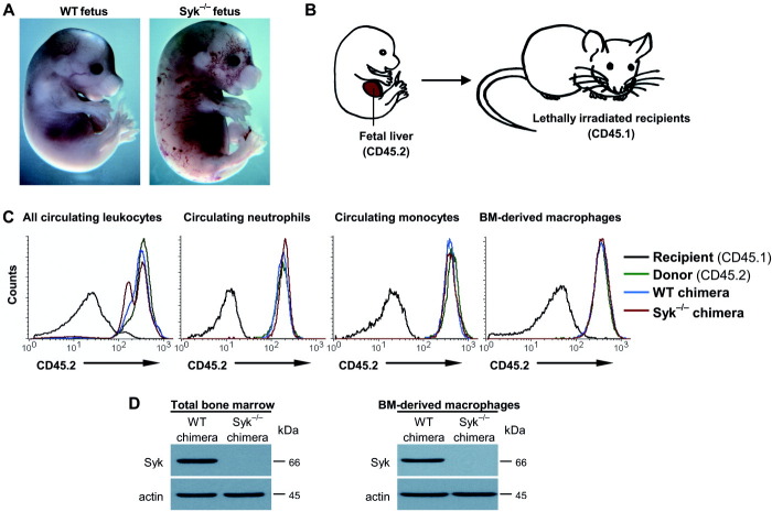
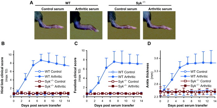
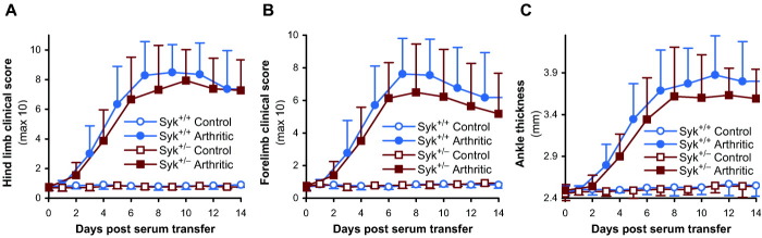
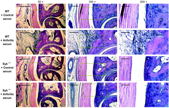
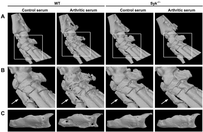
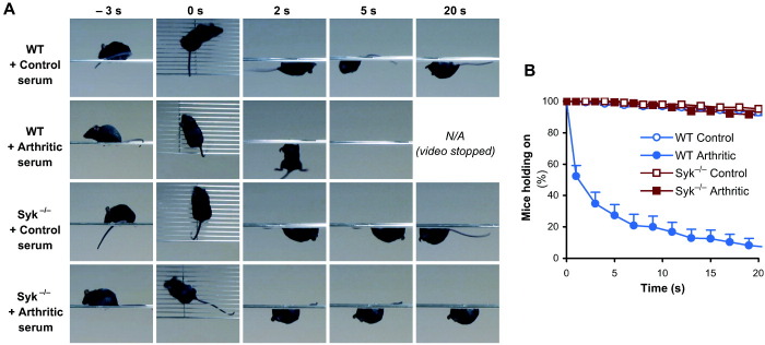
Methods: Syk(-/-) bone marrow chimeras carrying a Syk-deficient hematopoietic system were generated by transplanting Syk(-/-) fetal liver cells into lethally irradiated wild-type recipients. After complete repopulation of the hematopoietic compartment, autoantibody-mediated arthritis was induced by injection of arthritogenic K/BxN serum. Arthritis development was monitored by macroscopic and microscopic observation of the ankle joints, micro-computed tomography of bone morphology, as well as a joint function assay.

Results: Genetic deficiency of Syk in the hematopoietic compartment completely blocked the development of all macroscopic and microscopic signs of arthritis. The Syk(-/-) mutation also prevented the appearance of periarticular bone erosions. Finally, Syk(-/-) bone marrow chimeras were completely protected from arthritis-induced loss of articular function.

Conclusion: Our results indicate that Syk is critically involved in the development of all clinically relevant aspects of autoantibody-mediated K/BxN serum-transfer arthritis in experimental mice. These results provide the first in vivo genetic evidence of the role of Syk in the development of autoimmune arthritis.


Figures

Figure 1
Figure 1
Generation and analysis of Syk−/− bone marrow chimeric mice. A, Macroscopic appearance of wild-type (WT) and Syk−/− mouse fetuses at ∼17.5 days postcoitum (embryogenesis day 17.5). B, General scheme of fetal liver transplantation procedure. C, Flow cytometric analysis of donor marker (CD45.2) expression in all circulating leukocytes (CD45+ gate), circulating neutrophils (Gr-1+ gate), circulating monocytes (CD11b+Gr-1– gate), and bone marrow (BM)–derived macrophages (F4/80+ gate) from intact (nonchimeric) mice of the CD45.1-expressing recipient strain or the CD45.2-expressing donor (C57BL/6) strain, as well as from wild-type or Syk−/− bone marrow chimeras. D, Immunoblot analysis of Syk and β-actin protein levels in total bone marrow cells or bone marrow–derived macrophages. Results are representative of 3 or more independent experiments, each of which showed similar results. Actin was used as a loading control.
Figure 2
Figure 2
Assessment of the macroscopic signs of arthritis in Syk−/− bone marrow chimeric mice. A, Photographs of the hind limb of wild-type (WT) or Syk−/− chimeras 8 days after a single injection of 400 μl of arthritogenic (K/BxN) or control (BxN) serum. B–D, Quantification of arthritis severity by hind limb (B) or fore limb (C) clinical scoring or by measurement of ankle thickness (D). Experiments were performed on all 4 limbs of 6 control serum– and 11 arthritogenic serum–treated mice per genotype in 4 independent experiments. Shown are representative photographs (A) or the mean and SD values (B–D) of 2 limbs per mouse from all mice tested.
Figure 3
Figure 3
Normal arthritis development in heterozygous Syk+/− mice. Arthritis severity was quantified by hind limb (A) or fore limb (B) clinical scoring or by measurement of ankle thickness (C) in intact (nonchimeric) homozygous Syk+/+ or heterozygous Syk+/− mice after a single injection of 400 μl of arthritogenic (K/BxN) or control (BxN) serum. Experiments were performed on all 4 limbs of 5 control serum– and 10 arthritogenic serum–treated mice per genotype in 3 independent experiments. Values are the mean and SD of 2 limbs per mouse from all tested mice combined.
Figure 4
Figure 4
Histologic analysis of the ankle joints of Syk−/− bone marrow chimeric mice. Photomicrographs of hematoxylin and eosin–stained sagittal sections of the ankle joints of wild-type (WT) or Syk−/− chimeras 4 days after a single injection of 400 μl of arthritogenic (K/BxN) or control (BxN) serum are shown. In each case, sections obtained at 50× magnification show the joint formed between the tibia (above) and the astragalus (below) bones (the latter corresponding to the human talus), with the posterior surface pointing to the left. Boxed areas in images shown at 50× magnification are shown at 100× magnification in images in the center column, and boxed areas in images shown at 100× magnification are shown at 200× magnification in images in the right column. Bars = 500 μm, 200 μm, and 100 μm for 50×, 100×, and 200× magnification, respectively. Images are representative of a large number of sections obtained from 3 independent experiments, each of which showed similar results.
Figure 5
Figure 5
Micro–computed tomography (micro-CT) of the ankle joints of Syk−/− bone marrow chimeric mice. A and B, Three-dimensional reconstruction of the ankle bones of wild-type (WT) and Syk−/− bone marrow chimeras obtained by micro-CT at 9 μm voxel size on day 8 after a single injection of 400 μl of arthritogenic (K/BxN) or control (BxN) serum. Boxed areas in A are shown at higher digital magnification in B. Arrow in B indicates the first distal tarsal bone. C, Three-dimensional images of the first distal tarsal bone obtained by micro-CT at 5 μm voxel size after complete digestion of the ankle joints. Images are representative of 3 ankles per group obtained from 3 independent experiments, each of which showed similar results.
Figure 6
Figure 6
Protection of Syk−/− bone marrow chimeric mice from arthritis-induced loss of articular function. Wild-type (WT) or Syk−/− bone marrow chimeras were injected with a single dose of 400 μl of arthritogenic (K/BxN) or control (BxN) serum. On days 8–12 after serum injection, the mice were placed on a custom-made wire grid, which was then flipped over, and the length of time the mice held on to the wire grid during a 20-second assessment period was recorded. A, Snapshots from the video captures of mice of the indicated genotype and treatment groups 10 days after serum injection. B, Percentages of mice that were able to hold on to the grid at the indicated time points after the grid was flipped over. The experiment was repeated 15–20 times per mouse (n = 6 control serum–treated and n = 11 arthritic serum–treated chimeras per genotype in 4 independent experiments). Shown are representative snapshots (A) or the mean and SD values (B) for each group of mice tested.

Similar articles

Cited by

References

    1. Firestein GS. Evolving concepts of rheumatoid arthritis. Nature. 2003;423:356–61. - PubMed
    1. McInnes IB, Schett G. Cytokines in the pathogenesis of rheumatoid arthritis. Nat Rev Immunol. 2007;7:429–42. - PubMed
    1. Lundberg K, Nijenhuis S, Vossenaar ER, Palmblad K, van Venrooij WJ, Klareskog L, et al. Citrullinated proteins have increased immunogenicity and arthritogenicity and their presence in arthritic joints correlates with disease severity. Arthritis Res Ther. 2005;7:R458–67. - PMC - PubMed
    1. Kuhn KA, Kulik L, Tomooka B, Braschler KJ, Arend WP, Robinson WH, et al. Antibodies against citrullinated proteins enhance tissue injury in experimental autoimmune arthritis. J Clin Invest. 2006;116:961–73. - PMC - PubMed
    1. Edwards JC, Szczepanski L, Szechinski J, Filipowicz-Sosnowska A, Emery P, Close DR, et al. Efficacy of B-cell–targeted therapy with rituximab in patients with rheumatoid arthritis. N Engl J Med. 2004;350:2572–81. - PubMed

Publication types

MeSH terms