. 2010 Jun;62(6):1630-40.
doi: 10.1002/art.27439.

Autocitrullination of human peptidyl arginine deiminase type 4 regulates protein citrullination during cell activation

Affiliations

Autocitrullination of human peptidyl arginine deiminase type 4 regulates protein citrullination during cell activation

Felipe Andrade et al. Arthritis Rheum. 2010 Jun.

Abstract

Objective: To address mechanisms that control the activity of human peptidyl arginine deiminase type 4 (PAD-4).

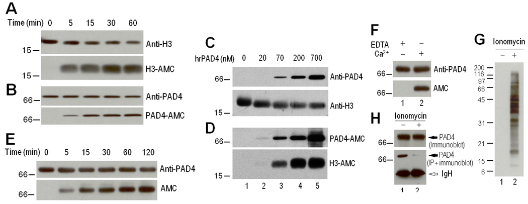
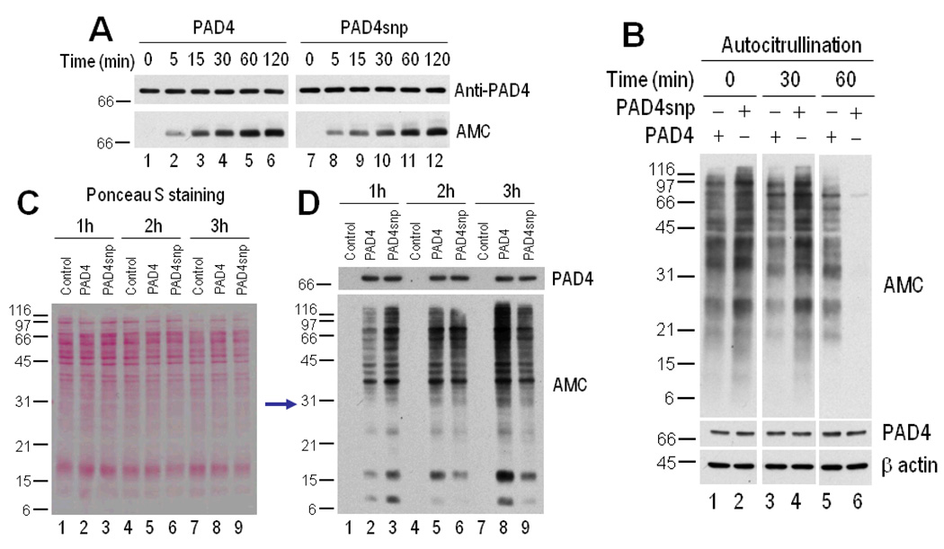
Methods: PAD-4 autocitrullination was determined by anti-modified citrulline immunoblotting, using purified recombinant and endogenous PAD-4 from activated human primary neutrophils and cell lines expressing PAD-4. The citrullination sites in PAD-4 were determined by mass spectrometry. Mechanisms of autocitrullination-induced inactivation and the functional consequences of autocitrullination in PAD-4 polymorphic variants were addressed using purified components and cell lines expressing PAD-4 wild-type, PAD-4 mutant, and PAD-4 polymorphic variants relevant to rheumatoid arthritis (RA).

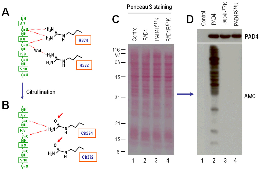
Results: PAD-4 is autocitrullinated in vitro and during activation of primary cells and cell lines expressing PAD-4. Interestingly, this modification inactivated the function of the enzyme. The efficiency of inactivation differed among genetically defined PAD-4 variants relevant to RA. PAD-4 was citrullinated at 10 sites, which are clustered into 3 distinct regions, including a cluster of arginines around the active site cleft where Arg-372 and -374 were identified as the potential autocitrullination targets that inactivate the enzyme. Autocitrullination also modified the structure of PAD-4, abrogating its recognition by multiple rabbit antibodies, but augmenting its recognition by human anti-PAD-4 autoantibodies.

Conclusion: Our findings suggest that autocitrullination regulates the production of citrullinated proteins during cell activation, and that this is affected by structural polymorphisms in PAD-4. Autocitrullination also influences PAD-4 structure and immune response.


Figures

Figure 1
Figure 1
Autocitrullination of recombinant human peptidyl arginine deiminase type 4 (rhPAD-4) in vitro. AE, Recombinant human PAD-4 (700 nM in A, B, and E or 0–700 nM in C and D) was coincubated with 700 nM human recombinant histone H3.1 or incubated alone (E) in the presence of 10 mM CaCl2 at 37°C for 0–60 minutes (A and B), for 60 minutes (C and D), or for 0–120 minutes (E). F, Recombinant human PAD-4 (700 nM) was incubated in the presence of 5 mM EDTA or 10 mM CaCl2 for 120 minutes at 37°C. After termination of the reactions, the samples were electrophoresed and noncitrullinated and citrullinated proteins were detected by immunoblotting with anti–histone H3, anti–PAD-4 C-terminal antibody, and anti–modified citrulline (AMC). G and H, Immunoprecipitation (IP) of PAD-4 from control and activated neutrophils. Primary human neutrophils in Hanks' balanced salt solution containing 2 mM CaCl2 were incubated in the absence (lane 1) or presence (lane 2) of 1 µM ionomycin for 2 hours at 37°C. Following the incubation, the cells were lysed in radioimmunoprecipitation assay buffer. The samples were directly electrophoresed (G and top panel of H) or used to immunoprecipitate endogenous PAD-4 using a rabbit anti-human PAD-4 C-terminal antibody (bottom panel of H). Protein citrullination was detected by anti–modified citrulline immunoblotting (G), and endogenous PAD-4 (top panel of H) or immunoprecipitated PAD-4 (bottom panel of H) was detected by immunoblotting using the rabbit anti–PAD-4 C-terminal antibody.
Figure 2
Figure 2
Autocitrullination of PAD-4 during cell activation. A, Immunoprecipitation of control and citrullinated rhPAD-4 by rabbit antibodies and by human rheumatoid arthritis (RA) sera. Noncitrullinated (native) rhPAD-4 and citrullinated (cit) rhPAD-4 were immunoprecipitated using rabbit anti-human PAD-4 antibodies against the C-terminal (Cterm) or N-terminal (Nterm) domains, or using human anti–PAD-4 RA sera (i.e., 2454, 1067, 2489, and 2314). Purified immune complexes were electrophoresed, and PAD-4 was detected by immunoblotting using anti–PAD-4 C-terminal antibody. B, Immunoprecipitation of PAD-4 from control and activated neutrophil lysates. Cell lysates were generated in radioimmunoprecipitation assay (RIPA) buffer from control neutrophils (lanes 1 and 3) and ionomycin-activated neutrophils (lanes 2 and 4), and endogenous PAD-4 was immunoprecipitated using human anti–PAD-4 serum. Then, control and ionomycin-activated samples were divided in two and immunoblotted using anti–PAD-4 C-terminal antibody (lanes 1 and 2) or anti–modified citrulline (lanes 3 and 4). C, Protein citrullination in 293T cells that were transiently transfected to express green fluorescent protein (GFP)–PAD-4. After 48 hours, the cells were incubated in the absence (lane 1) or presence (lane 2) of 1 µM ionomycin for 2 hours at 37°C. Following the incubation, the cells were lysed in RIPA buffer and electrophoresed to detect protein citrullination by anti–modified citrulline. D, Immunoprecipitation of GFP–PAD-4 from control and activated 293T-transfected cells. Purified immune complexes were electrophoresed, and equal protein loading was visualized by ponceau S staining (top) prior to anti–modified citrulline immunoblotting (bottom). See Figure 1 for other definitions.
Figure 3
Figure 3
Autocitrullination of PAD-4 inhibits its function. A, Generation of noncitrullinated rhPAD-4 (lane 1) and citrullinated rhPAD-4 (lane 2). Recombinant human PAD-4 was incubated in the absence or presence of 10 mM CaCl2 for 120 minutes at 37°C. Samples were immunoblotted with rabbit anti–PAD-4 (top) and anti–modified citrulline (bottom). B, Citrullination activity of control and citrullinated rhPAD-4. HL-60 cell lysates were incubated with buffer alone (lanes 1 and 4), noncitrullinated rhPAD-4 (lanes 2 and 5), or citrullinated (cit) rhPAD-4 (lanes 3 and 6) for 15 minutes (lanes 1–3) or 60 minutes (lanes 4–6) at 37°C. After termination of the reactions, the samples were electrophoresed and immunoblotted using anti–modified citrulline, anti–PAD-4 C-terminal antibody, and anti–β-actin (loading control). See Figure 1 for other definitions.
Figure 4
Figure 4
Clustering of the potential citrullination sites in peptidyl arginine deiminase type 4 (PAD-4) into 3 distinct regions. A, Schematic representation of the secondary structure of PAD-4 (adapted from ref.24). The secondary structure elements in the N-terminal and C-terminal domains are shown in blue and yellow, respectively. Bars show -helices; arrows show β strands; and broken lines show disordered regions. The potential arginine targets for citrullination are shown in red. B, Tertiary structure of PAD-4, revealing clustering of citrullination sites into 3 distinct regions. The cluster comprising Arg-372, -374, -383, and -394 spans the active site cleft. The N-terminal and C-terminal domains are shown in blue and yellow, respectively, while the sites of arginine deimination are indicated in red. The model shown was generated from the Molecular Modeling Database (National Center for Biotechnology Information), according to coordinates generated by Arita et al (16). Adapted by permission from Macmillan Publishers Ltd: Nat Struct Mol Biol 2004;11(8):777–83. Copyright 2004.
Figure 5
Figure 5
The PAD-4 citrullination sites Arg-372 and Arg-374 regulate PAD-4 activity during cell activation. A, Schematic diagram of the interactions between Arg-374 and Arg-372 at the active site cleft of PAD-4 and N-terminal residues in histone H3-1 (adapted from ref.26). Arg-374 makes multiple hydrogen bonds with backbone carbonyl oxygens of the Ala-7 (A7) and Arg-8 (R8) residues in histone H3.1 (green), and Arg-372 recognizes the carbonyl oxygen of the Arg-8 residue by means of water (Wat)–mediated hydrogen bonds. B, Conversion of Arg-372 and Arg-374 to citrullines (Cit) during PAD-4 autocitrullination, potentially distressing the interactions shown in A. C and D, The PAD4 mutants PAD4R372K and PAD4R374K lack citrullination activity. The 293T cells were mock transfected (lane 1) or transiently transfected to express PAD-4 (lane 2), PAD-4R372K (lane 3), or PAD-4R374K (lane 4). At 48 hours posttransfection, the cells were stimulated with 1 εM ionomycin for 1 hour. After termination of the reactions, the samples were electrophoresed, and equal protein loading was visualized by ponceau S staining (C) prior to anti–modified citrulline immunoblotting (bottom panel of D). PAD-4 expression was visualized by immunoblotting (top panel of D). See Figure 1 for other definitions.
Figure 6
Figure 6
PAD-4 variants have distinct patterns of inactivation induced by autocitrullination. A, Autocitrullination of rhPAD-4 and rhPAD-4snp variants. Recombinant human PAD-4 and rhPAD-4snp were incubated in the presence of 10 mM CaCl2 at 37°C for 0–120 minutes. Noncitrullinated and citrullinated proteins were detected by immunoblotting with anti–PAD-4 (top) and anti–modified citrulline (bottom), respectively. B, Citrullination activity of control and citrullinated rhPAD-4 and rhPAD-4snp. Recombinant human PAD-4 (lanes 1, 3, and 5) or rhPAD-4snp (lanes 2, 4, and 6) were incubated in the presence of 10 mM CaCl2 for 0, 30, or 60 minutes at 37°C, and further incubated with HL-60 cell lysate for an additional 15 minutes at 37°C. After termination of the reactions, the samples were electrophoresed and immunoblotted using anti–modified citrulline, anti–PAD-4, and anti–β-actin (loading control). C and D, Citrullination activity of PAD-4 and PAD-4snp during cell activation. The 293T cells were mock transfected (lanes 1, 4, and 7) or transiently transfected to express PAD-4 (lanes 2, 5, and 8) or PAD-4snp (lanes 3, 6, and 9). At 48 hours posttransfection, the cells were stimulated with 1 µM ionomycin for 1 hour (lanes 1–3), 2 hours (lanes 4–6), 3 hours (lanes 7–9), and 5–6 hours (results not shown). After termination of the reactions, the samples were electrophoresed, and equal protein loading was visualized by ponceau S staining (C) prior to anti–modified citrulline immunoblotting (bottom panel of D). PAD-4 expression was visualized by immunoblotting (top panel of D). See Figure 1 for other definitions.

Similar articles

Cited by

References

    1. Gelato KA, Fischle W. Role of histone modifications in defining chromatin structure and function. Biol Chem. 2008;389(4):353–363. - PubMed
    1. Zhang Q, Wang Y. High mobility group proteins and their post-translational modifications. Biochim Biophys Acta. 2008;1784(9):1159–1166. - PMC - PubMed
    1. Tarcsa E, Marekov LN, Mei G, Melino G, Lee SC, Steinert PM. Protein unfolding by peptidylarginine deiminase. Substrate specificity and structural relationships of the natural substrates trichohyalin and filaggrin. J Biol Chem. 1996;271(48):30709–30716. - PubMed
    1. Doyle HA, Mamula MJ. Posttranslational modifications of self-antigens. Ann N Y Acad Sci. 2005;1050:1–9. - PubMed
    1. Rogers GE. Occurrence of citrulline in proteins. Nature. 1962;194:1149–1151. - PubMed

Publication types