

Long-term consequences of Sox9 depletion on inner ear development
- PMID: 20201105
- PMCID: PMC2847000
- DOI: 10.1002/dvdy.22259
Long-term consequences of Sox9 depletion on inner ear development
Abstract
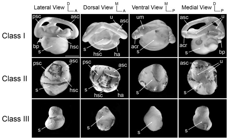
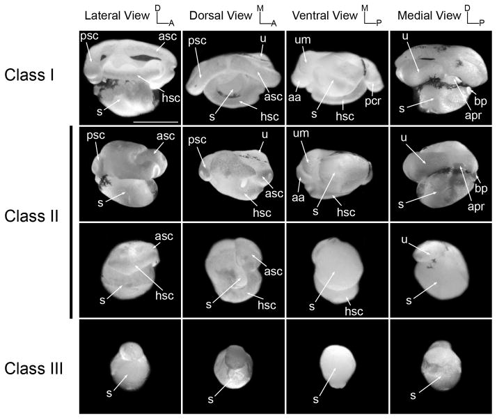
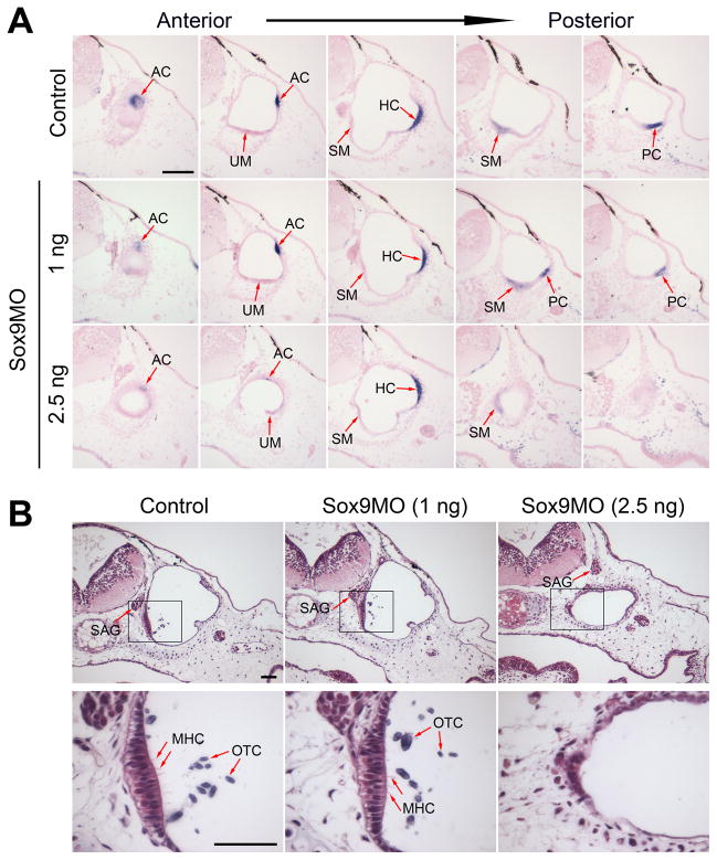
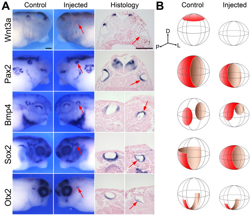
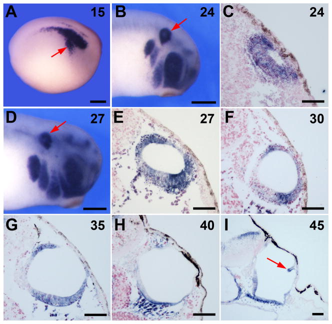
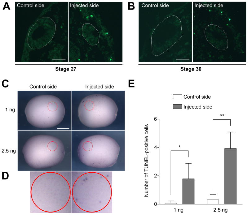
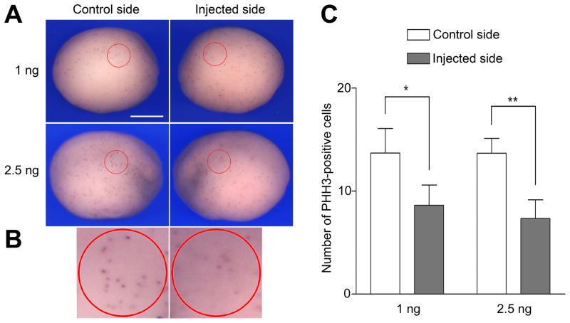
The transcription factor Sox9 has been implicated in inner ear formation in several species. To investigate the long-term consequences of Sox9 depletion on inner ear development we analyzed the inner ear architecture of Sox9-depleted Xenopus tadpoles generated by injection of increasing amounts of Sox9 morpholino antisense oligonucleotides. We found that Sox9-depletion resulted in major defects in the development of vestibular structures, semicircular canals and utricle, while the ventrally located saccule was less severely affected in these embryos. Consistent with this phenotype, we observed a specific loss of the dorsal expression of Wnt3a expression in the otic vesicle of Sox9 morphants, associated with an increase in cell death and a reduction in cell proliferation in the region of the presumptive otic epithelium. We propose that, in addition to its early role in placode specification, Sox9 is also required for the maintenance of progenitors in the otic epithelium.
Figures
Similar articles
-
Specification of the otic placode depends on Sox9 function in Xenopus.Development. 2004 Apr;131(8):1755-63. doi: 10.1242/dev.01066. Epub 2004 Mar 17. Development. 2004. PMID: 15084460
-
A restrictive role for Hedgehog signalling during otic specification in Xenopus.Dev Biol. 2003 Aug 15;260(2):325-38. doi: 10.1016/s0012-1606(03)00242-2. Dev Biol. 2003. PMID: 12921735
-
The role of Paraxial Protocadherin in Xenopus otic placode development.Biochem Biophys Res Commun. 2006 Jun 23;345(1):239-47. doi: 10.1016/j.bbrc.2006.04.068. Epub 2006 Apr 25. Biochem Biophys Res Commun. 2006. PMID: 16678122
-
Changing shape and shaping change: Inducing the inner ear.Semin Cell Dev Biol. 2017 May;65:39-46. doi: 10.1016/j.semcdb.2016.10.006. Epub 2016 Oct 29. Semin Cell Dev Biol. 2017. PMID: 27989562 Review.
-
Signaling regulating inner ear development: cell fate determination, patterning, morphogenesis, and defects.Congenit Anom (Kyoto). 2015 Feb;55(1):17-25. doi: 10.1111/cga.12072. Congenit Anom (Kyoto). 2015. PMID: 25040109 Review.
Cited by
-
Six1 proteins with human branchio-oto-renal mutations differentially affect cranial gene expression and otic development.Dis Model Mech. 2020 Mar 3;13(3):dmm043489. doi: 10.1242/dmm.043489. Dis Model Mech. 2020. PMID: 31980437 Free PMC article.
-
SOX9 and SOX10 control fluid homeostasis in the inner ear for hearing through independent and cooperative mechanisms.Proc Natl Acad Sci U S A. 2022 Nov 16;119(46):e2122121119. doi: 10.1073/pnas.2122121119. Epub 2022 Nov 7. Proc Natl Acad Sci U S A. 2022. PMID: 36343245 Free PMC article.
-
Exome sequencing of families from Ghana reveals known and candidate hearing impairment genes.Commun Biol. 2022 Apr 19;5(1):369. doi: 10.1038/s42003-022-03326-8. Commun Biol. 2022. PMID: 35440622 Free PMC article.
-
Loss of Mst1/2 activity promotes non-mitotic hair cell generation in the neonatal organ of Corti.NPJ Regen Med. 2022 Oct 25;7(1):64. doi: 10.1038/s41536-022-00261-4. NPJ Regen Med. 2022. PMID: 36280668 Free PMC article.
-
Six1 and Irx1 have reciprocal interactions during cranial placode and otic vesicle formation.Dev Biol. 2019 Feb 1;446(1):68-79. doi: 10.1016/j.ydbio.2018.12.003. Epub 2018 Dec 6. Dev Biol. 2019. PMID: 30529252 Free PMC article.
References
-
- Bagheri-Fam S, Barrionuevo F, Dohrmann U, Günther T, Schüle R, Kemler R, Mallo M, Kanzler B, Scherer G. Long-range upstream and downstream enhancers control distinct subsets of the complex spatiotemporal Sox9 expression pattern. Dev Biol. 2006;291:382–397. - PubMed
-
- Baird RA, Torres MA, Schuff NR. Hair cell regeneration in bullfrog vestibular otolith organs following aminoglycoside toxicity. Hear Res. 1993;65:164–174. - PubMed
-
- Barrionuevo F, Bagheri-Fam S, Klattig J, Kist R, Taketo MM, Englert C, Scherer G. Homozygous inactivation of Sox9 causes complete XY sex reversal in mice. Biol Reprod. 2006;74:195–201. - PubMed
-
- Barrionuevo F, Naumann A, Bagheri-Fam S, Speth V, Taketo MM, Scherer G, Neubüser A. Sox9 is required for invagination of the otic placode in mice. Dev Biol. 2008;317:213–224. - PubMed
-
- Bernard P, Tang P, Liu S, Dewing P, Harley VR, Vilain E. Dimerization of SOX9 is required for chondrogenesis, but not for sex determination. Hum Mol Genet. 2003;12:1755–1765. - PubMed
Publication types
MeSH terms
Substances
Grants and funding
LinkOut - more resources
Full Text Sources
Research Materials
