. 2010 Mar;86(3):194-204.
doi: 10.3109/09553000903418603.

DNA damage intensity in fibroblasts in a 3-dimensional collagen matrix correlates with the Bragg curve energy distribution of a high LET particle

Affiliations

DNA damage intensity in fibroblasts in a 3-dimensional collagen matrix correlates with the Bragg curve energy distribution of a high LET particle

Andres I Roig et al. Int J Radiat Biol. 2010 Mar.

Abstract

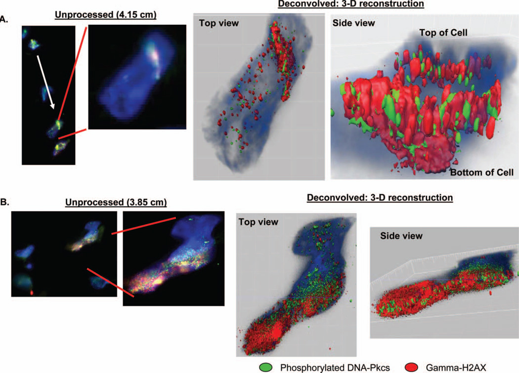
Purpose: The DNA double-strand break (DSB) damage response induced by high energy charged particles on lung fibroblast cells embedded in a 3-dimensional (3-D) collagen tissue equivalents was investigated using antibodies to the DNA damage response proteins gamma-histone 2AX (gamma-H2AX) and phosphorylated DNA-PKcs (p-DNA-PKcs).

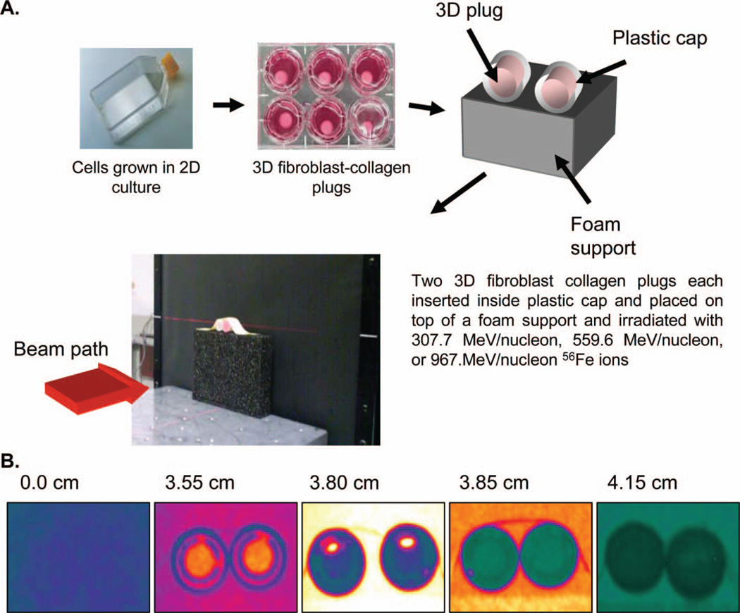
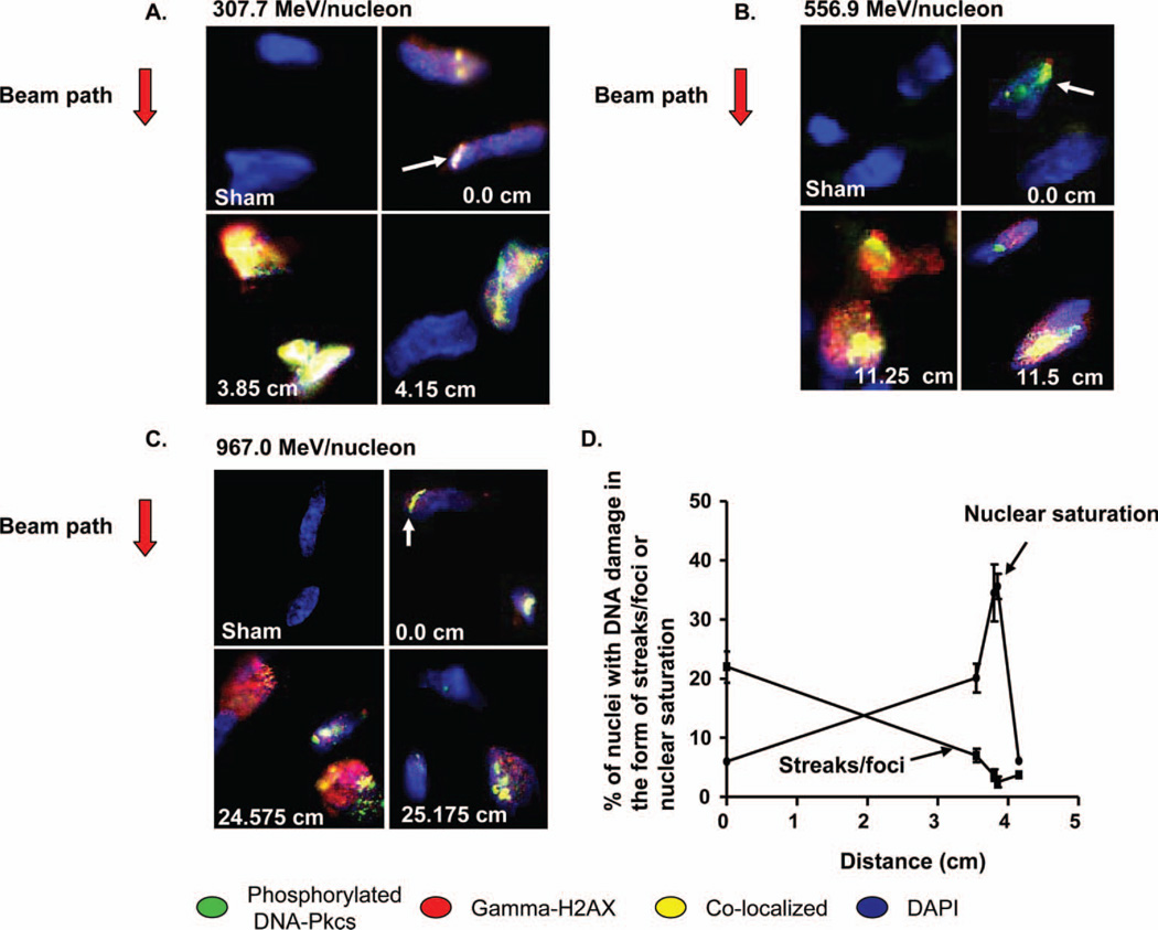
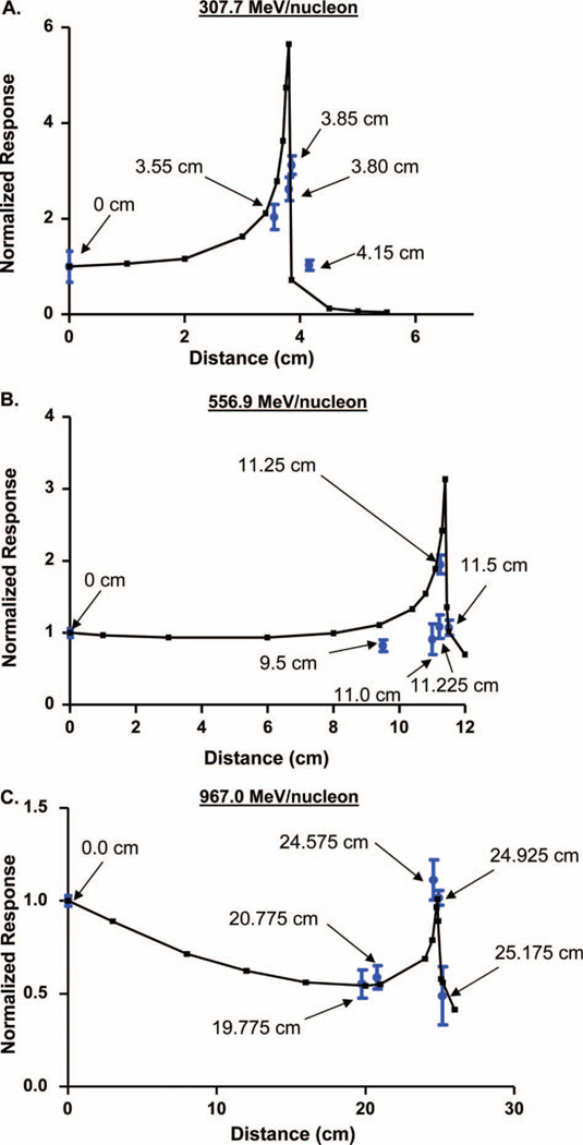
Materials and methods: 3-D tissue equivalents were irradiated in positions across the linear distribution of the Bragg curve profiles of 307.7 MeV/nucleon, 556.9 MeV/nucleon, or 967.0 MeV/nucleon (56)Fe ions at a dose of 0.30 Gy.

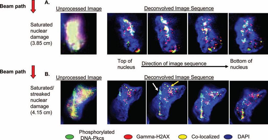
Results: Patterns of discrete DNA damage streaks across nuclei or saturated nuclear damage were observed, with saturated nuclear damage being more predominant as samples were positioned closer to the physical Bragg peak. Quantification of the DNA damage signal intensities at each distance for each of the examined energies revealed a biological Bragg curve profile with a pattern of DNA damage intensity similar to the physical Bragg curve for the particular energy. Deconvolution microscopy of nuclei with streaked or saturated nuclear damage pattern revealed more details of the damage, with evidence of double-strand breaks radially distributed from the main particle track as well as multiple discrete tracks within saturated damage nuclei.

Conclusions: These 3-D culture systems can be used as a biological substrate to better understand the interaction of heavy charged particles of different energies with tissue and could serve as a basis to model space-radiation-induced cancer initiation and progression.


Conflict of interest statement

Declaration of interest: The authors do not have any conflict of interests or financial arrangements to disclose.

Figures

Figure 1
Figure 1
(A) Collagen-fibroblast plugs are made from lung fibroblast cells grown in conventional monolayer. Once mixed with collagen to make a three dimensional tissue equivalent, they are properly oriented for exposure to high LET radiation at the NASA Space Radiation Laboratory (NSRL) at Brookhaven National Laboratory (BNL). (B) Placement of plugs at different positions in the Bragg curve of a 307.7 MeV/nucleon 56Fe particle shows varying energies deposited to tissues depending on their location in the Bragg curve.
Figure 2
Figure 2
(A), (B), (C) DNA damage in nuclei of specimens placed at various locations along the Bragg peak for 307.7 MeV/nucleon, 556.9 MeV/nucleon, or 967.0 MeV/nucleon 56Fe ions, respectively. The beam direction for each energy is indicated next to the left of each panel. The damage is appreciated as either discrete damage streaks or foci (white arrows) or saturated nuclear damage. All images taken at 40× objective magnification. (D) Percentage of nuclei in a 307.7 MeV/nucleon specimen showing streaks/foci or nuclear saturation damage. Co-localised γ-H2AX and DNA-PKcs nuclear streak damage is more prevalent in distances farther away from the Bragg peak while saturated nuclear damage is more common closer to the peak. Error bars indicate the standard error for the mean (SEM) for three separate counting experiments.
Figure 3
Figure 3
(A), (B), (C) Normalised DNA damage intensities calculated by averaging the mean grey scale values of the DNA damage intensities corresponding to 56Fe ions with 307.7 MeV/nucleon, 556.9 MeV/nucleon, or 967.0 MeV/nucleon energy super-imposed over the physical Bragg profiles for each particle energy. Four separate intensity measuring experiments (between 80 and 100 total cell nuclei) were performed for each spatial location in the Bragg curve for each particular energy as well for non-irradiated controls. Error bars indicate the standard error for the mean (SEM) for these four separate intensity measuring experiments.
Figure 4
Figure 4
Magnified pictures of representative cell nuclei from specimens with saturated nuclear damage or mixed streaked- saturated nuclear damage for a 307.7 MeV/nucleon 56Fe beam. Both images come from samples sectioned parallel to the beam path. (A). Close up view of the saturated nuclear damage pattern processed with an epifluorescent microscope and corresponding deconvolved image sequences of the saturated damage revealing distinct particle tracks at multiple optical sections. Unprocessed image obtained at 40× objective magnification. Deconvolved images obtained at 60× objective magnification. (B). Sample nucleus with mixed streaked-saturated damage. Deconvolution shows a particle track with small tangential secondary tracks. Unprocessed image obtained at 60× magnification. Deconvolved images obtained at 60× objective magnification.
Figure 5
Figure 5
3D reconstructions of deconvolved images from nuclei with streaked (panel A; sample sectioned parallel to beam path, white arrow illustrating direction of particle path) and nuclear saturation damage (panel B; sample sectioned transverse to the particle path, particle enters cell head-on). Left-most unprocessed epifluorescent images obtained at 40× objective magnification while right-sided unprocessed images were obtained at 60× objective magnification. 3-D reconstructions (right-most images, top and side views of the nuclei) demonstrate a surface rendition of the γ-H2AX and DNA-PKcs damage.

Similar articles

Cited by

References

    1. Asaithamby A, Uematsu N, Chatterjee A, Story MD, Burma S, Chen DJ. Repair of HZE-particle-induced DNA double-strand breaks in normal human fibroblasts. Radiation Research. 2008;169:437–446. - PubMed
    1. Bekker-Jensen S, Lukas C, Kitagawa R, Melander F, Kastan MB, Bartek J, Lukas J. Spatial organization of the mammalian genome surveillance machinery in response to DNA strand breaks. The Journal of Cell Biology. 2006;173:195–206. - PMC - PubMed
    1. Chan DW, Chen BP, Prithivirajsingh S, Kurimasa A, Story MD, Qin J, Chen DJ. Autophosphorylation of the DNA-dependent protein kinase catalytic subunit is required for rejoining of DNA double-strand breaks. Genes and Development. 2002;16:2333–2338. - PMC - PubMed
    1. Chen BP, Chan DW, Kobayashi J, Burma S, Asaithamby A, Morotomi-Yano K, Botvinick E, Qin J, Chen DJ. Cell cycle dependence of DNA-dependent protein kinase phosphorylation in response to DNA double strand breaks. Journal of Biological Chemistry. 2005;280:14709–14715. - PubMed
    1. Cowell IG, Sunter NJ, Singh PB, Austin CA, Durkacz BW, Tilby MJ. gammaH2AX foci form preferentially in euchromatin after ionising-radiation. PLoS ONE. 2007;2:e1057. - PMC - PubMed

Publication types

MeSH terms

LinkOut - more resources