Review
. 2010 Apr;8(3):240-52.
doi: 10.2174/157016210791111124.

The Vpu protein: new concepts in virus release and CD4 down-modulation

Affiliations
Review

The Vpu protein: new concepts in virus release and CD4 down-modulation

Autumn Ruiz et al. Curr HIV Res. 2010 Apr.

Abstract

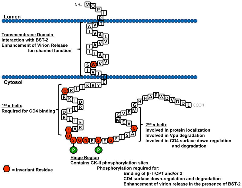
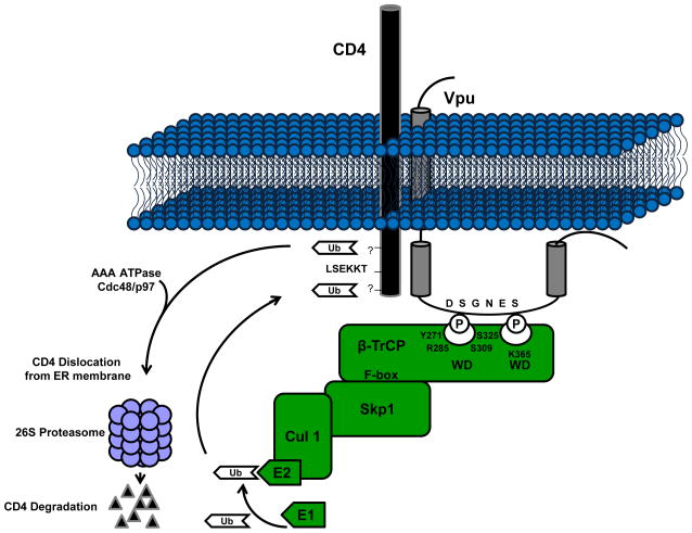
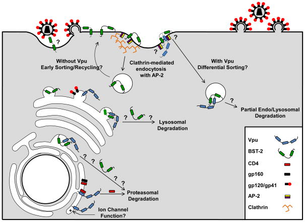
Human immunodeficiency virus type 1 (HIV-1) and several simian immunodeficiency viruses (SIV) encode for a transmembrane protein known as Vpu (viral protein U). While one of the smallest of the HIV-1 proteins, it has two important functions within virus-infected cells. The first of these functions is the down-regulation of the CD4 receptor to prevent its interaction with the HIV-1 envelope glycoprotein. Vpu interacts with the CD4 receptor in the rough endoplasmic reticulum (RER), resulting in its re-translocation across the RER and subsequent degradation via the proteasomal pathway. The second major function of the Vpu protein is to facilitate release of virus from infected cells. Previous studies have shown that virus release is cell type specific, suggesting that certain cells may express a restriction factor that inhibits virus release in the absence of Vpu. Recently, bone marrow stromal antigen 2 (BST-2/HM1.24/CD317/tetherin) has been identified as this factor. This review will focus on new findings within the last four years on the role of Vpu in CD4 down-regulation and the restriction of virus release from cells. We will relate these findings to virus pathogenesis and propose questions regarding BST-2 as a restriction factor.


Figures

Fig. 1
Fig. 1
Schematic diagram of the membrane orientation of the consensus subtype B Vpu protein (strain HXB2 with a corrected methionine at the N-terminus). The amino acids within the hexagons represent amino acids that were found to be invariant among all vpu subtypes (50)
Fig. 2
Fig. 2
Down-modulation of cell surface CD4 expression by Vpu. In the rough endoplasmic reticulum, Vpu binds CD4 through an interaction between the first α-helix within the cytoplasmic domain of Vpu and a specific motif within the cytoplasmic domain of CD4 (LSEKKT). Vpu is phosphorylated by several different isoforms of casein kinase II. This phosphorylation recruits and binds the cellular F-box protein β-TrCP. CD4 is then ubiquitinated via the SCFβ-TrCP E3 Ub ligase complex after which it is shunted to the proteasome for degradation through an unknown mechanism.
Fig. 3
Fig. 3
Possible mechanisms by which Vpu enhances virion release from cells. Vpu allows efficient transport of Env proteins to the site of virion assembly. Vpu is synthesized on the rough endoplasmic reticulum and binds CD4 releasing gp160 for processing and cleavage into gp120/gp41 which may be transported to the site of assembly. Vpu may function as a viroporin. Vpu resides within the rough endoplasmic reticulum, trans-Golgi network and at the cell surface. It is unknown whether the ability of Vpu to serve as an ion channel functions to enhance virion release at any of these intracellular locations. Vpu removes BST-2 from the site of virion assembly. In the absence of Vpu, BST-2 is expressed at the cell surface. The orientation used by BST-2 to “tether” virions to the surface remains unknown, however, the GPI anchor resides within lipid rafts where HIV-1 virions assemble and bud. This potentiates a model where the N-terminal transmembrane remains anchored within the cell membrane while the C-terminal GPI anchor is embedded in the virion membrane. BST-2 is internalized from lipid rafts on the cell surface by clathrin-mediated endocytosis. Two tyrosine residues within the N-terminal cytoplasmic tail are required for an interaction with a Δα-adaptin of the AP-2 complex. It is possible that in the presence of Vpu the targeting of endocytosed BST-2 containing vesicles is altered for endo-lysosomal degradation. Vpu may also potentially interact with BST-2 in trans-Golgi network and target it for lysosomal or proteasomal degradation.

Similar articles

Cited by

References

    1. Barlow KL, Ajao AO, Clewley JP. Journal of Virology. 2003;77:6879–6888. - PMC - PubMed
    1. Bartee E, McCormack A, Früh K. PLoS Pathogens. 2006;10:e107. - PMC - PubMed
    1. Besnard-Guerin C, Belaidouni N, Lassot I, Segeral E, Jobart A, Marchal C, Benarous R. Journal of Biological Chemistry. 2004;279:788–795. - PubMed
    1. Binette J, Dube M, Mercier J, Halawani D, Latterich M, Cohen EA. Retrovirology. 2007;4:75. - PMC - PubMed
    1. Bour S, Schubert U, Strebel K. Journal of Virology. 1995;69:1510–1520. - PMC - PubMed

Publication types

MeSH terms

Substances