

Extracellular signal-regulated kinase 1/2 (ERK1/2) signaling in cardiac hypertrophy
- PMID: 20201891
- PMCID: PMC5941943
- DOI: 10.1111/j.1749-6632.2009.05088.x
Extracellular signal-regulated kinase 1/2 (ERK1/2) signaling in cardiac hypertrophy
Abstract
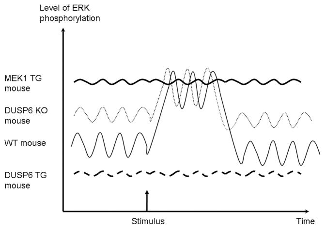
Cardiac hypertrophy results from increased mechanical load on the heart and through the action of neurohumoral mediators. ERK1/2 are known to be activated in response to almost every stress- and agonist-induced hypertrophic stimulus examined to date, suggesting the straightforward hypothesis that these kinases are required for promoting the cardiac growth response. However, recent data from genetically modified mouse models suggest a more complicated picture. For example, inducible expression of dual-specificity phosphatase 6, an ERK1/2-inactivating phosphatase, eliminated ERK1/2 phosphorylation in transgenic mice, but it did not diminish the hypertrophic response to pressure overload. Similarly, Erk1-/- and Erk2+/- mice showed no reduction in stimulus-induced cardiac growth in vivo. However, blockade or deletion of cardiac ERK1/2 did predispose the heart to decompensation and failure after long-term pressure overload. Thus, ERK1/2 signaling is not to be absolutely necessary for mediating cardiac hypertrophy, although it does appear to provide critical protective effects/signals during stress-stimulation.
Figures
References
-
- HO KK, PINSKY JL, KANNEL WB, LEVY D. The epidemiology of heart failure: the Framingham Study. J Am Coll Cardiol. 1993;22:6A–13A. - PubMed
-
- KLEIN L, et al. Pharmacologic therapy for patients with chronic heart failure and reduced systolic function: review of trials and practical considerations. Am J Cardiol. 2003;91:18F–40F. - PubMed
-
- BUENO OF, MOLKENTIN JD. Involvement of extracellular signal-regulated kinases 1/2 in cardiac hypertrophy and cell death. Circ Res. 2002;91:776–81. - PubMed
-
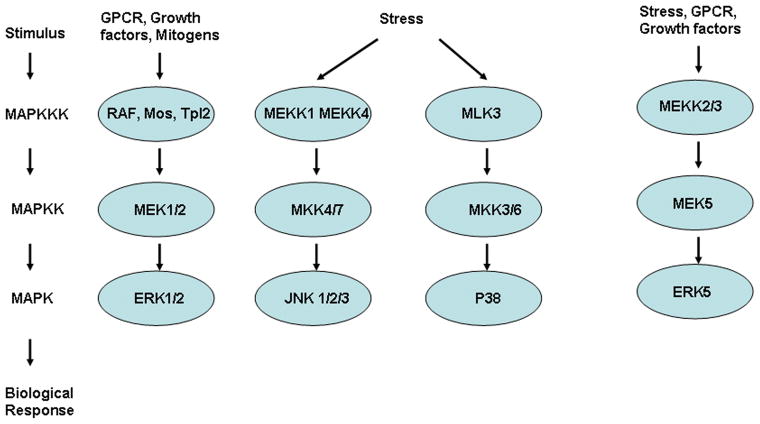
- GARRINGTON TP, JOHNSON GL. Organization and regulation of mitogen-activated protein kinase signaling pathways. Curr Opin Cell Biol. 1999;11:211–8. - PubMed
-
- WELLBROCK C, KARASARIDES M, MARAIS R. The RAF proteins take centre stage. Nat Rev Mol Cell Biol. 2004;5:875–85. - PubMed
Publication types
MeSH terms
Substances
Grants and funding
LinkOut - more resources
Full Text Sources
Miscellaneous
