

The role of mechanical forces in the torsional component of cardiac looping
- PMID: 20201892
- PMCID: PMC2837544
- DOI: 10.1111/j.1749-6632.2009.05089.x
The role of mechanical forces in the torsional component of cardiac looping
Abstract
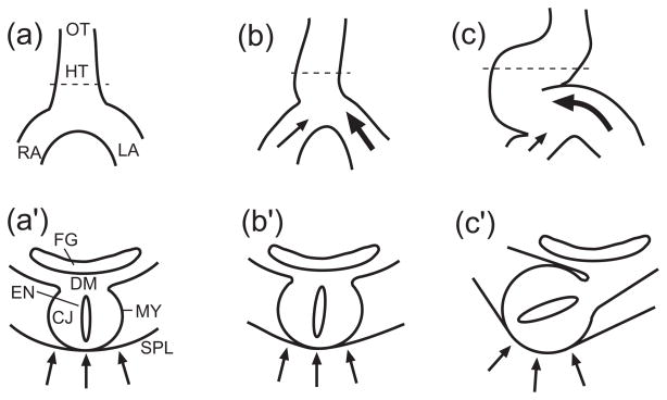
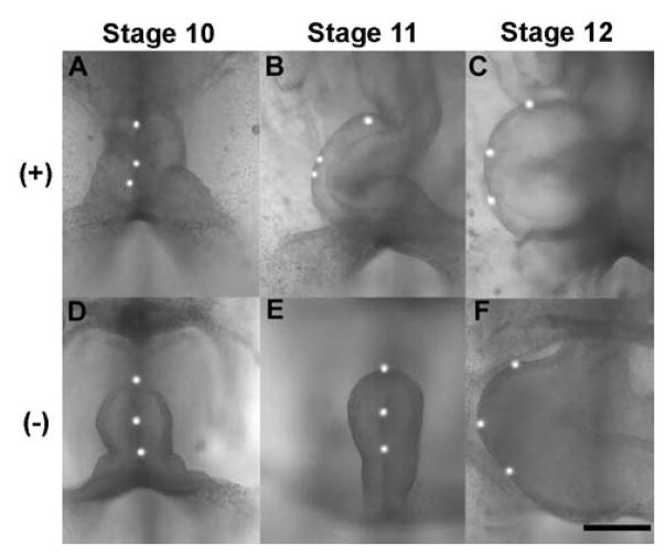
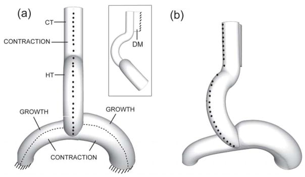
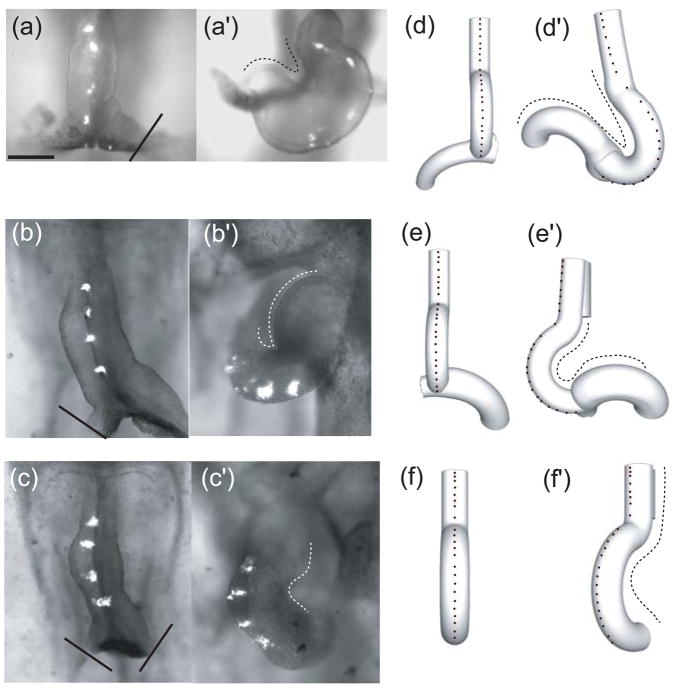
During early development, the initially straight heart tube (HT) bends and twists (loops) into a curved tube to lay out the basic plan of the mature heart. The physical mechanisms that drive and regulate looping are not yet completely understood. This paper reviews our recent studies of the mechanics of cardiac torsion during the first phase of looping (c-looping). Experiments and computational modeling show that torsion is primarily caused by forces exerted on the HT by the primitive atria and the splanchnopleure, a membrane that presses against the ventral surface of the heart. Experimental and numerical results are described and integrated to propose a hypothesis for cardiac torsion, and key aspects of our hypothesis are tested using experiments that perturb normal looping. For each perturbation, the models predict the correct qualitative response. These studies provide new insight into the mechanisms that drive and regulate cardiac looping.
Figures
Similar articles
-
On modeling morphogenesis of the looping heart following mechanical perturbations.J Biomech Eng. 2008 Dec;130(6):061018. doi: 10.1115/1.2978990. J Biomech Eng. 2008. PMID: 19045547 Free PMC article.
-
On the role of intrinsic and extrinsic forces in early cardiac S-looping.Dev Dyn. 2013 Jul;242(7):801-16. doi: 10.1002/dvdy.23968. Epub 2013 Jun 5. Dev Dyn. 2013. PMID: 23553909 Free PMC article.
-
The role of mechanical forces in dextral rotation during cardiac looping in the chick embryo.Dev Biol. 2004 Aug 15;272(2):339-50. doi: 10.1016/j.ydbio.2004.04.033. Dev Biol. 2004. PMID: 15282152
-
Biophysical mechanisms of cardiac looping.Int J Dev Biol. 2006;50(2-3):323-32. doi: 10.1387/ijdb.052045lt. Int J Dev Biol. 2006. PMID: 16479500 Review.
-
Cardiac looping in the chick embryo: a morphological review with special reference to terminological and biomechanical aspects of the looping process.Anat Rec. 2000 Jul 1;259(3):248-62. doi: 10.1002/1097-0185(20000701)259:3<248::AID-AR30>3.0.CO;2-K. Anat Rec. 2000. PMID: 10861359 Review.
Cited by
-
Why is cytoskeletal contraction required for cardiac fusion before but not after looping begins?Phys Biol. 2015 Jan 30;12(1):016012. doi: 10.1088/1478-3975/12/1/016012. Phys Biol. 2015. PMID: 25635663 Free PMC article.
-
Bending and twisting the embryonic heart: a computational model for c-looping based on realistic geometry.Front Physiol. 2014 Aug 12;5:297. doi: 10.3389/fphys.2014.00297. eCollection 2014. Front Physiol. 2014. PMID: 25161623 Free PMC article.
-
Zebrafish as a Versatile Model for Cardiovascular Research: Peering into the Heart of the Matter.Cells. 2025 Apr 2;14(7):531. doi: 10.3390/cells14070531. Cells. 2025. PMID: 40214485 Free PMC article. Review.
-
Cell chirality in cardiovascular development and disease.APL Bioeng. 2020 Aug 25;4(3):031503. doi: 10.1063/5.0014424. eCollection 2020 Sep. APL Bioeng. 2020. PMID: 32903894 Free PMC article. Review.
-
A right-handed signalling pathway drives heart looping in vertebrates.Nature. 2017 Sep 6;549(7670):86-90. doi: 10.1038/nature23454. Nature. 2017. PMID: 28880281 Free PMC article.
References
-
- Harvey RP. Cardiac looping --- an uneasy deal with laterality. Semin Cell Dev Biol. 1998;9:101–108. - PubMed
-
- Srivastava D, Olson EN. Knowing in your heart what’s right. Trends Cell Biol. 1997;7:447–453. - PubMed
-
- Taber LA. Biophysical mechanisms of cardiac looping. Int J Dev Biol. 2006;50:323–332. - PubMed
-
- DeHaan RL. Development of form in the embryonic heart. An experimental approach. Circulation. 1967;35:821–833. - PubMed
-
- Hamburger V, Hamilton HL. A series of normal stages in the development of the chick embryo. J Morphol. 1951;88:49–92. - PubMed
Publication types
MeSH terms
Grants and funding
LinkOut - more resources
Full Text Sources
