

L-type calcium channel as a cardiac oxygen sensor
- PMID: 20201898
- PMCID: PMC3064564
- DOI: 10.1111/j.1749-6632.2009.05095.x
L-type calcium channel as a cardiac oxygen sensor
Abstract
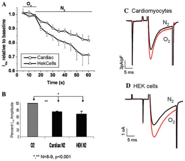
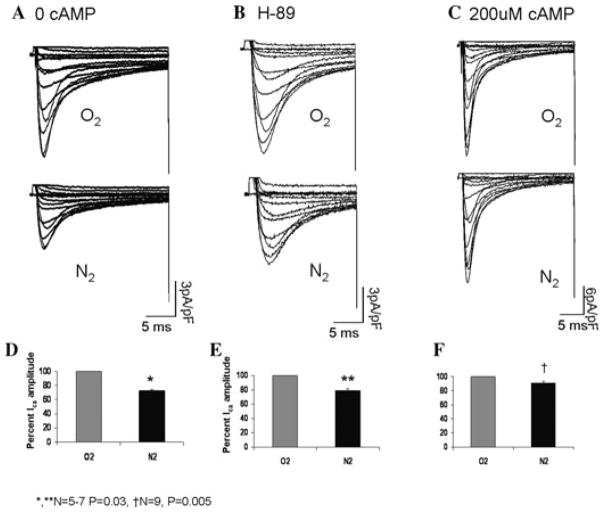
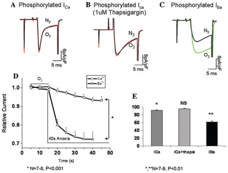
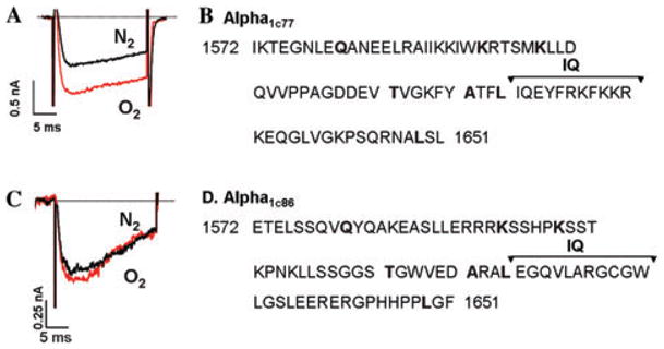
Acute oxygen sensing in the heart is thought to occur through redox regulation and phosphorylation of membrane channels. Here we report a novel O2-sensing mechanism involving the C-terminus of the L-type Ca2+ channel and regulated by PKA phosphorylation. In patch-clamped myocytes, oxygen deprivation decreased ICa within 40 s. The suppressive effect of anoxia was relieved by PKA-mediated phosphorylation only when Ca2+ was the charge carrier, whereas phosphorylated IBa remained sensitive to O2 withdrawal. Suppression of Ca2+ release by thapsigargin did not alter the response of ICa to anoxia, suggesting a mandatory role for Ca2+ influx and not Ca2+-induced Ca2+ release (CICR) in O2 regulation of the channel. Consistent with this idea, mutation of 80 amino acids in the Ca2+/CaM-binding domain of the recombinant alpha1C subunit that removes Ca2+ dependent inactivation (CDI) abolished O2 sensitivity of the channel. Our findings suggest that the Ca2+/CaM binding domain of the L-type Ca2+ may represent a molecular site for O2 sensing of the heart.
Conflict of interest statement
The authors declare no conflicts of interest.
Figures
Similar articles
-
Hypoxic regulation of cardiac Ca2+ channel: possible role of haem oxygenase.J Physiol. 2012 Sep 1;590(17):4223-37. doi: 10.1113/jphysiol.2012.236570. Epub 2012 Jul 2. J Physiol. 2012. PMID: 22753548 Free PMC article.
-
C terminus L-type Ca2+ channel calmodulin-binding domains are 'auto-agonist' ligands in rabbit ventricular myocytes.J Physiol. 2003 Aug 1;550(Pt 3):731-8. doi: 10.1113/jphysiol.2003.043778. Epub 2003 Jun 13. J Physiol. 2003. Retraction in: J Physiol. 2015 May 1;593(9):2245. doi: 10.1113/JP270443. PMID: 12807986 Free PMC article. Retracted.
-
Modulation of Ca2+ signalling in rat atrial myocytes: possible role of the alpha1C carboxyl terminal.J Physiol. 2003 Oct 15;552(Pt 2):437-47. doi: 10.1113/jphysiol.2003.048330. J Physiol. 2003. PMID: 14561827 Free PMC article.
-
New aspects in cardiac L-type Ca2+ channel regulation.Biochem Soc Trans. 2020 Feb 28;48(1):39-49. doi: 10.1042/BST20190229. Biochem Soc Trans. 2020. PMID: 32065210 Review.
-
Regulation of calcium slow channels of heart by cyclic nucleotides and effects of ischemia.Adv Pharmacol. 1994;31:1-24. doi: 10.1016/s1054-3589(08)60605-5. Adv Pharmacol. 1994. PMID: 7873407 Review.
Cited by
-
Hypoxic regulation of cardiac Ca2+ channel: possible role of haem oxygenase.J Physiol. 2012 Sep 1;590(17):4223-37. doi: 10.1113/jphysiol.2012.236570. Epub 2012 Jul 2. J Physiol. 2012. PMID: 22753548 Free PMC article.
-
Regulation of Ca2+ signaling by acute hypoxia and acidosis in rat neonatal cardiomyocytes.J Mol Cell Cardiol. 2018 Jan;114:58-71. doi: 10.1016/j.yjmcc.2017.10.004. Epub 2017 Oct 12. J Mol Cell Cardiol. 2018. PMID: 29032102 Free PMC article.
-
Hypoxia. 4. Hypoxia and ion channel function.Am J Physiol Cell Physiol. 2011 May;300(5):C951-67. doi: 10.1152/ajpcell.00512.2010. Epub 2010 Dec 22. Am J Physiol Cell Physiol. 2011. PMID: 21178108 Free PMC article. Review.
-
Aggravation of myocardial dysfunction by injurious mechanical ventilation in LPS-induced pneumonia in rats.Respir Res. 2013 Sep 18;14(1):92. doi: 10.1186/1465-9921-14-92. Respir Res. 2013. PMID: 24047433 Free PMC article.
-
Regulation of Ca2+ signaling by acute hypoxia and acidosis in cardiomyocytes derived from human induced pluripotent stem cells.Cell Calcium. 2019 Mar;78:1-14. doi: 10.1016/j.ceca.2018.12.006. Epub 2018 Dec 12. Cell Calcium. 2019. PMID: 30579812 Free PMC article.
References
-
- Schneider MF, Chandler WK. Voltage dependent charge movement of skeletal muscle: a possible step in excitation-contraction coupling. Nature. 1973;242:244–246. - PubMed
-
- Morad M, Soldatov N. Calcium channel inactivation: possible role in signal transduction and Ca2+ signaling. Cell Calcium. 2005;38:223–231. - PubMed
-
- Vittone L, et al. Time course and mechanisms of phosphorylation of phospholamban residues in ischemia-reperfused rat hearts: dissociation of phospholamban phosphorylation pathways. J Mol Cell Cardiol. 2002;34:39–50. - PubMed
-
- Yamaoka K, Kameyama M. Regulation of L-type Ca2+ channels in the heart: overview of recent advances. Mol Cell Biochem. 2003;253:3–13. - PubMed
MeSH terms
Substances
Grants and funding
LinkOut - more resources
Full Text Sources
Miscellaneous
