

Developmental basis of adult cardiovascular diseases: valvular heart diseases
- PMID: 20201901
- PMCID: PMC3371607
- DOI: 10.1111/j.1749-6632.2009.05098.x
Developmental basis of adult cardiovascular diseases: valvular heart diseases
Abstract
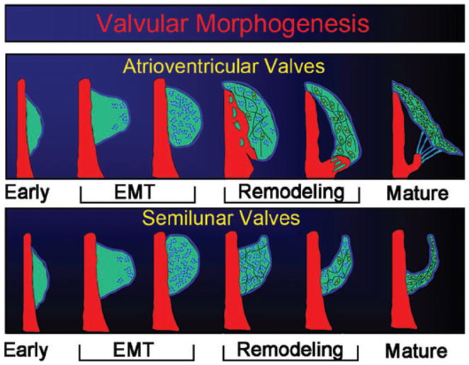
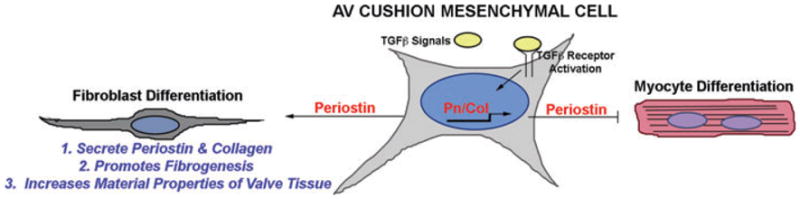
In this chapter, we review the working hypothesis that the roots of adult valvular heart disease (VHD) lie in embryonic development. Valvulogenesis is a complex process in which growth factors signal the process of endocardium-to-mesenchyme transformation (EMT) resulting in formation of prevalvular "cushions." The post-EMT processes, whereby cushions are morphogenetically remolded into valve leaflets, are less well understood, but they require periostin. Mice with targeted deletion of periostin develop degenerative changes similar to human forms of VHD. Mitral valves are also abnormally elongated in hypertrophic cardiomyopathy (HCM), which plays an important role in clinical disease expression. However, the mechanism for this is unclear, but correlates with enhanced expression of periostin in a specific population of ventricular cells derived from the embryonic proepicardial organ, which accumulate at sites where valvular endocardial EMT is reactivated. Collectively, these findings suggest that developmental mechanisms underlie adult valve responses to genetic mutations in degenerative VHD and HCM.
Conflict of interest statement
Figures
References
-
- Person AD, Klewer SE, Runyan RB. Cell biology of cardiac cushion development. Int Rev Cytol. 2005;243:287–335. - PubMed
-
- DeRuiter MC, Poelmann RE, VanMunsteren JC, et al. Embryonic endothelial cells transdifferentiate into mesenchymal cells expressing smooth muscle actins in vivo and in vitro. Circ Res. 1997;80:444–451. - PubMed
-
- Lincoln J, Alfieri CM, Yutzey KE. Development of heart valve leaflets and supporting apparatus in chicken and mouse embryos. Dev Dyn. 2004;230:239–250. - PubMed
Publication types
MeSH terms
Grants and funding
LinkOut - more resources
Full Text Sources
Medical
