. 2010 May;130(1):137-49.
doi: 10.1111/j.1365-2567.2010.03220.x. Epub 2010 Feb 26.

Optimal stimulation for CD70 induction on human monocyte-derived dendritic cells and the importance of CD70 in naive CD4(+) T-cell differentiation

Affiliations

Optimal stimulation for CD70 induction on human monocyte-derived dendritic cells and the importance of CD70 in naive CD4(+) T-cell differentiation

Kazue Arimoto-Miyamoto et al. Immunology. 2010 May.

Abstract

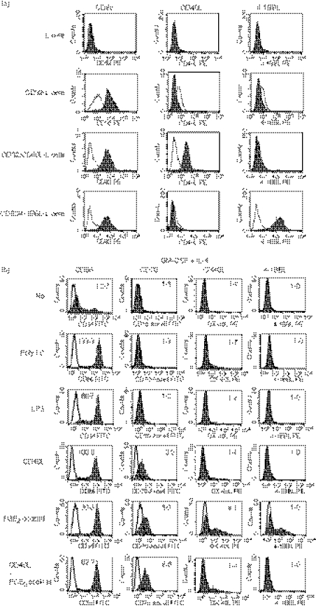
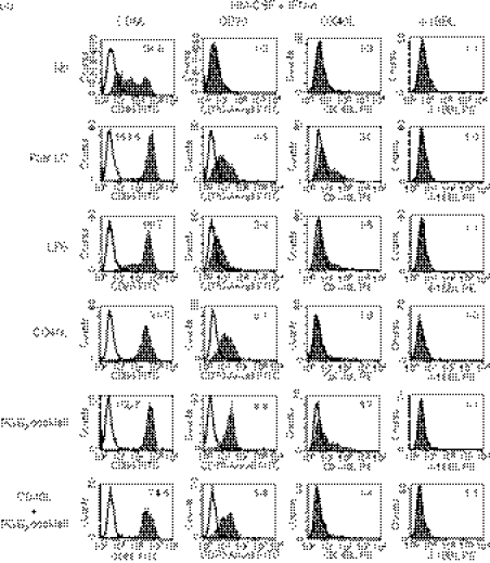
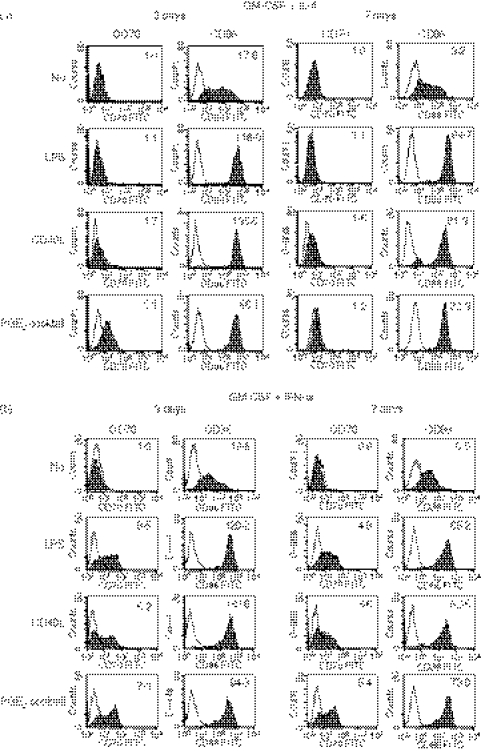
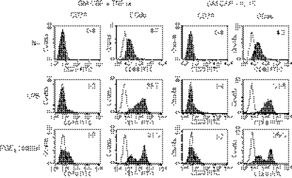
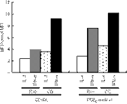
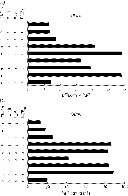
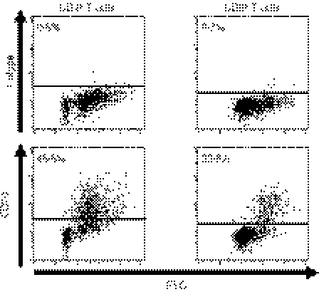
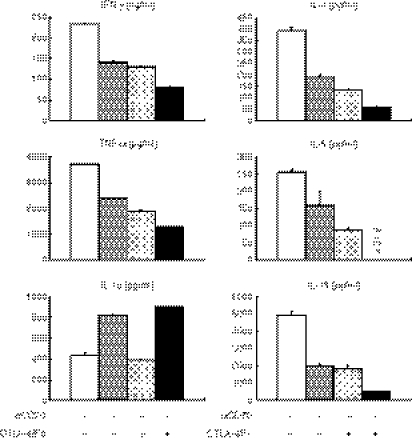
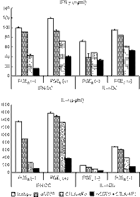
Studies in mice have shown that CD70 on dendritic cells (DCs) is sufficient to convert T-cell tolerance into immunity and hence induce anti-tumour immune responses. Therefore, it is important to investigate (i) optimal stimuli to induce CD70 on human monocyte-derived DCs (MoDCs), which are widely used for tumour immunotherapy, and (ii) the role of CD70 in functional differentiation of naive CD4(+) and CD8(+) T cells stimulated with MoDCs. We show that interferon-alpha (IFN-alpha) is a key cytokine to differentiate monocytes into DCs with the capacity to express CD70 upon maturation. CD70 expression on IFN-alpha-induced MoDCs was elicited by different categories of maturation-inducing factors (Toll-like receptor ligands, CD40 ligand and pro-inflammatory mediators), among which prostaglandin E(2) was most effective. Naive T cells stimulated with MoDCs also expressed CD70. Stimulation with MoDCs promoted naive CD4(+) T cells to acquire the ability to produce T helper type 1 and 2 cytokines in a CD70-dependent manner. In contrast, the CD70-CD27 interaction diminished the production of an immunoregulatory cytokine IL-10. The CD27 signal did not play a dominant role in the induction of effector molecules in naive CD8(+) T cells during the stimulation with MoDCs. This study adds a novel function to the versatile cytokines, type I IFNs, that is, the induction of CD70 on MoDCs. CD70 promotes naive CD4(+) T cells to acquire immunostimulatory activity through the DC-T-cell and T-cell-T-cell interactions during the stimulation with MoDCs. Hence, the CD70-CD27 interaction may play an important role in inducing effective immune responses in DC-based immunotherapy.


Figures

Figure 1
Figure 1
Expression of CD70, OX40 ligand (OX40L), 4-1BBL and CD86 on monocyte-derived dendritic cells (MoDCs) cultured in different conditions. (a) Parental L cells and those transfected with CD32 (CD32-L), CD32 and CD40L (CD32/CD40L-L) or CD32 and 4-1BBL (CD32/4-1BBL-L) were stained with phycoerythrin (PE) -labelled anti-CD32, anti-CD40L or anti-4-1BBL monoclonal antibody (mAb). Open histograms represent cells stained with isotype-matched control mAbs. (b, c) Monocytes were cultured for 3 days in R10 in the presence of interleukin-4 (IL-4) (b) or interferon-α (IFN-α) (c) together with granulocyte–macrophage colony-stimulating factor (GM-CSF) to induce immature IL-4-DCs or IFN-DCs, respectively. Thereafter, they were cultured for 3 days in the absence or presence of maturation-inducing stimuli [poly I:C, lipopolysaccharide (LPS), CD40L-L cells, or the prostaglandin E2 (PGE2) cocktail]. The cells were stained with fluorescein isothiocyanate (FITC) -labelled anti-CD86, anti-CD70 (clone BU69), PE-labelled anti-OX40L, or anti-4-1BBL mAb. Open histograms represent cells stained with isotype-matched control mAbs. The numbers shown with each histogram represent [mean fluorescence intensity (MFI) of each costimulatory molecule]/(MFI of isotype-matched control]. Data are representative of three experiments.
Figure 1
Figure 1
Expression of CD70, OX40 ligand (OX40L), 4-1BBL and CD86 on monocyte-derived dendritic cells (MoDCs) cultured in different conditions. (a) Parental L cells and those transfected with CD32 (CD32-L), CD32 and CD40L (CD32/CD40L-L) or CD32 and 4-1BBL (CD32/4-1BBL-L) were stained with phycoerythrin (PE) -labelled anti-CD32, anti-CD40L or anti-4-1BBL monoclonal antibody (mAb). Open histograms represent cells stained with isotype-matched control mAbs. (b, c) Monocytes were cultured for 3 days in R10 in the presence of interleukin-4 (IL-4) (b) or interferon-α (IFN-α) (c) together with granulocyte–macrophage colony-stimulating factor (GM-CSF) to induce immature IL-4-DCs or IFN-DCs, respectively. Thereafter, they were cultured for 3 days in the absence or presence of maturation-inducing stimuli [poly I:C, lipopolysaccharide (LPS), CD40L-L cells, or the prostaglandin E2 (PGE2) cocktail]. The cells were stained with fluorescein isothiocyanate (FITC) -labelled anti-CD86, anti-CD70 (clone BU69), PE-labelled anti-OX40L, or anti-4-1BBL mAb. Open histograms represent cells stained with isotype-matched control mAbs. The numbers shown with each histogram represent [mean fluorescence intensity (MFI) of each costimulatory molecule]/(MFI of isotype-matched control]. Data are representative of three experiments.
Figure 2
Figure 2
A short-term culture is sufficient to induce maximal levels of CD70 expression on monocyte-derived dendritic cells (MoDCs). Monocytes were cultured for 3 or 7 days in R10 in the presence of interleukin-4 (IL-4) (a) or interferon-α (IFN-α) (b) together with granulocyte–macrophage colony-stimulating factor (GM-CSF) to induce immature IL-4-DCs or IFN-DCs, respectively. Thereafter, the DCs were cultured for 3 days in the absence or presence of maturation-inducing stimuli [lipopolysaccharide (LPS), CD40L-L cells, or the prostaglandin E2 (PGE2) cocktail]. The cells were stained with fluorescein isothiocyanate (FITC) -labelled anti-CD70 or anti-CD86 monoclonal antibody (mAb). Open histograms represent cells stained with isotype-matched control mAbs. The numbers shown with each histogram represent [mean fluorescence intensity (MFI) of each costimulatory molecule]/(MFI of isotype-matched control). Data are representative of three experiments.
Figure 3
Figure 3
Tumour necrosis factor-α (TNF-α) or interleukin-15 (IL-15) is not efficient to induce the expression of CD70 on monocyte-derived dendritic cells (MoDCs). Monocytes were cultured for 7 days in R10 in the presence of TNF-α or IL-15 together with granulocyte–macrophage colony-stimulating factor (GM-CSF) to induce immature TNF-DCs or IL-15-DCs, respectively. Thereafter, they were cultured for 3 days in the absence or presence of maturation-inducing stimuli (LPS or the PGE2 cocktail). The cells were stained with fluorescein isothiocyanate (FITC) -labelled anti-CD70 or anti-CD86 monoclonal antibody (mAb). Open histograms represent cells stained with isotype-matched control mAbs. The numbers shown with each histogram represent [mean fluorescence intensity (MFI) of each costimulatory molecule]/(MFI of isotype-matched control). Data are representative of two experiments. Because there were many dead cells when TNF-DCs and IL-15-DCs were stimulated with CD40L-transfected L cells, these data are not shown. When monocytes were cultured for 3 days to induce immature DCs, similar data were obtained.
Figure 4
Figure 4
Serum-free CellGro DC Medium is efficient to induce the expression of CD70 on monocyte-derived dendritic cells (MoDCs). Monocytes were cultured for 3 days in R10 or CellGro DC Medium (CG) in the presence of interleukin-4 (IL-4) or interferon-α (IFN-α) together with granulocyte–macrophage colony-stimulating factor (GM-CSF) to induce immature DCs. Thereafter, they were stimulated with CD40L-L cells or the prostaglandin E2 (PGE2) cocktail for 3 days in the same medium used for the first 3 days. The cells were stained with fluorescein isothiocyanate (FITC) -labelled anti-CD70 or anti-CD86 monoclonal antibody. Data are shown by [mean fluorescence intensity (MFI) of CD70]/(MFI of isotype-matched control). Data are representative of three experiments.
Figure 5
Figure 5
Tumour necrosis factor-α (TNF-α), interleukin-1β (IL-1β), plus prostaglandin E2 (PGE2) induce full expression of CD70. Immature interferon-treated dendritic cells (IFN-DCs) were cultured for 3 days in the absence or presence of the indicated combinations of components of the PGE2 cocktail in R10. Expression levels of CD70 (a) and CD86 (b) are shown by [mean fluorescence intensity (MFI) of each costimulatory molecule]/(MFI of isotype-matched control). Data are representative of three experiments.
Figure 6
Figure 6
Naive CD4+ and CD8+ T cells gain the expression of CD70 after stimulation with dendritic cells (DCs). Purified CD4+ CD45RA+ T cells or CD8+ CD45RA+ CCR7+ T cells were cocultured for 8 days with interferon-DCs stimulated with the prostaglandin E2 cocktail. The T cells were harvested and stained with fluorescein isothiocyanate-labelled anti-CD70 monoclonal antibody, and were analysed by flow cytometry. The percentages of CD70+ cells are indicated on the plot. Data are representative of three experiments.
Figure 7
Figure 7
The CD70–CD27 interaction promotes naive CD4+ T cells to acquire the ability to produce immunostimulatory cytokines while prevents them from producing interleukin-10 (IL-10) during the stimulation with CD70+ monocyte-derived dendritic cell s(MoDCs). CD4+ CD45RA+ T cells were cocultured for 8 days with allogeneic interferon-DCs stimulated with the prostaglandin E2 cocktail in the presence of 10 μg/ml anti-CD70 monoclonal antibody (mAb), 5 μg/ml human CTLA-4/Fc chimeric protein, or 10 μg/ml isotype-matched control mAb. The stimulated T cells were harvested, and were restimulated at 1 × 10⁶ cells/ml with plate-bound 10 μg/ml anti-CD3 and 1 μg/ml soluble anti-CD28 mAbs for 24 h. The supernatants were harvested and analysed for cytokines using enzyme-linked immunosorbent assay. Error bars indicate standard deviation of duplicate measurements. Data are representative of four experiments.
Figure 8
Figure 8
The CD70–CD27 signal enhances the production of T helper type 1 and 2 cytokines by naive CD4+ T cells irrespective of the expression of CD70 on dendritic cells (DCs) before coculture. CD4+ CD45RA+ T cells were cocultured for 8 days with the following allogeneic monocyte-derived (Mo) DCs: (i) interferon (IFN) -DCs stimulated with tumour necrosis factor-α (TNF-α), interleukin-1β (IL-1β), and IL-6, (ii) IFN-DCs stimulated with the prostaglandin E2 (PGE2) cocktail, (iii) IL-4-DCs stimulated with TNF-α, IL-1β and IL-6, or (iv) IL-4-DCs stimulated with the PGE2 cocktail, in the presence of 10 μg/ml anti-CD70 monoclonal antibody (mAb), 5 μg/ml human CTLA-4/Fc chimeric protein, or 10 μg/ml isotype-matched control mAb. The stimulated T cells were harvested, and were restimulated at 1 × 10⁶ cells/ml with plate-bound 10 μg/ml anti-CD3 and 1 μg/ml soluble anti-CD28 mAbs for 24 h. The supernatants were harvested and analysed for cytokine by enzyme-linked immunosorbent assay. Error bars indicate standard deviation of duplicate measurements. Data are representative of two experiments.

Similar articles

Cited by

References

    1. Watts TH. TNF/TNFR family members in costimulation of T cell responses. Annu Rev Immunol. 2005;23:23–68. - PubMed
    1. Borst J, Hendriks J, Xiao Y. CD27 and CD70 in T cell and B cell activation. Curr Opin Immunol. 2005;17:275–81. - PubMed
    1. Hendriks J, Gravestein LA, Tesselaar K, van Lier RAW, Schumacher TNM, Borst J. CD27 is required for generation and long-term maintenance of T cell immunity. Nat Immunol. 2000;1:433. - PubMed
    1. Hendriks J, Xiao Y, Borst J. CD27 promotes survival of activated T cells and complements CD28 in generation and establishment of the effector T cell pool. J Exp Med. 2003;198:1369–80. - PMC - PubMed
    1. Arens R, Tesselaar K, Baars PA, et al. Constitutive CD27/CD70 interaction induces expansion of effector-type T cells and results in IFN-γ-mediated B cell depletion. Immunity. 2001;15:801. - PubMed

Publication types

MeSH terms

Substances

LinkOut - more resources