Comparative Study
. 2010 May;113(4):871-80.
doi: 10.1111/j.1471-4159.2010.06653.x. Epub 2010 Feb 24.

Hemopressins and other hemoglobin-derived peptides in mouse brain: comparison between brain, blood, and heart peptidome and regulation in Cpefat/fat mice

Affiliations
Comparative Study

Hemopressins and other hemoglobin-derived peptides in mouse brain: comparison between brain, blood, and heart peptidome and regulation in Cpefat/fat mice

Julia S Gelman et al. J Neurochem. 2010 May.

Abstract

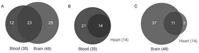
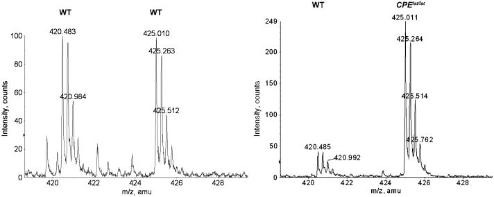
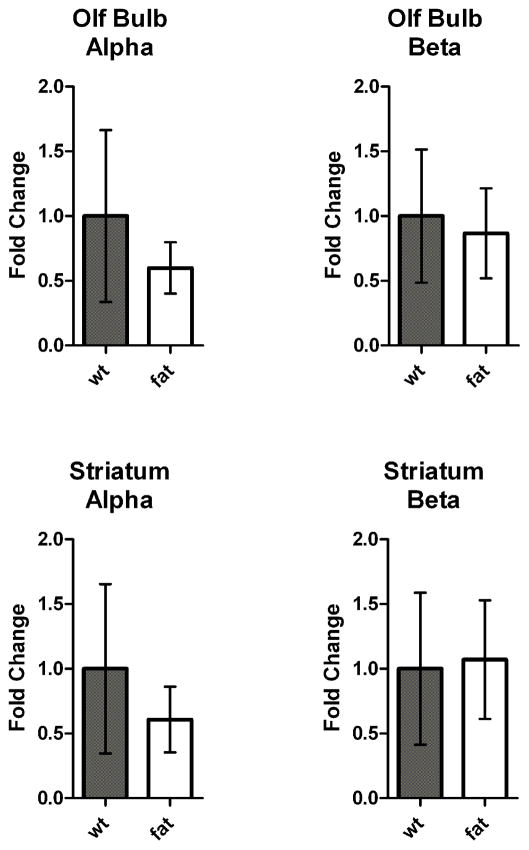
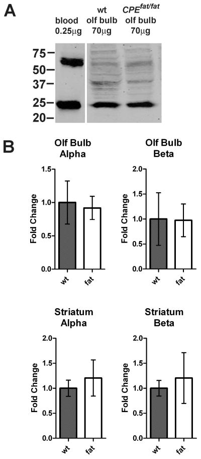
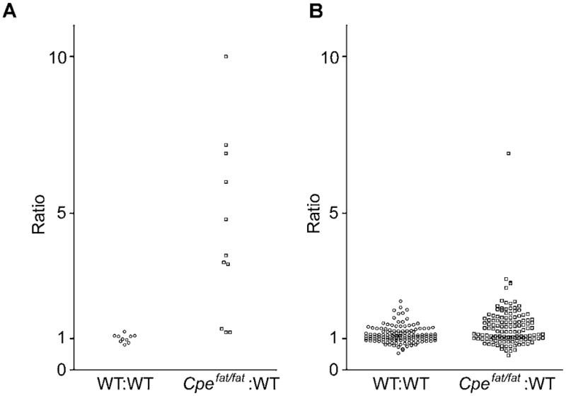
Many hemoglobin-derived peptides are present in mouse brain, and several of these have bioactive properties including the hemopressins, a related series of peptides that bind to cannabinoid CB1 receptors. Although hemoglobin is a major component of red blood cells, it is also present in neurons and glia. To examine whether the hemoglobin-derived peptides in brain are similar to those present in blood and heart, we used a peptidomics approach involving mass spectrometry. Many hemoglobin-derived peptides are found only in brain and not in blood, whereas all hemoglobin-derived peptides found in heart were also seen in blood. Thus, it is likely that the majority of the hemoglobin-derived peptides detected in brain are produced from brain hemoglobin and not erythrocytes. We also examined if the hemopressins and other major hemoglobin-derived peptides were regulated in the Cpe(fat/fat) mouse; previously these mice were reported to have elevated levels of several hemoglobin-derived peptides. Many, but not all of the hemoglobin-derived peptides were elevated in several brain regions of the Cpe(fat/fat) mouse. Taken together, these findings suggest that the post-translational processing of alpha and beta hemoglobin into the hemopressins, as well as other peptides, is up-regulated in some but not all Cpe(fat/fat) mouse brain regions.


Figures

Figure 1
Figure 1
Comparison of hemoglobin derived peptidome between tissues. A. Hemoglobin-derived peptides found in blood versus those found in brain, overlap of circles distinguishes those peptides that were seen in both blood and brain. Comparison of blood and heart is in panel B and brain versus heart is in panel C.
Figure 2
Figure 2
Representative spectra showing levels of peptides in wild-type and Cpefat/fat mice. Left panel: ions at m/z 420.483 and 425.010 (monoisotopic peaks) correspond to mono-D0- and D9- TMAB labeled RVD-hemopressin-α in olfactory bulb in an experiment evaluating WT vs WT mice. Ratio of D0 to D9 is 1.0, illustrating no change in peptide level in WT mice. Right panel: ions at 420.485 and 425.011 (monoisotopic peaks) correspond to mono-D0- and D9-TMAB labeled RVD-hemopressin-α in olfactory bulb in an experiment evaluating Cpefat/fat mice vs WT mice. Quantification of this spectra illustrates RVD-hemopressin-α is 5.5 fold higher in the Cpefat/fat mouse sample as compared to the WT mouse sample.
Figure 3
Figure 3
Quantitative real-time PCR of hemoglobin mRNA levels in wild-type and Cpefat/fat mice. Results are shown as the fold-change in expression. Fold change was calculated using the ΔΔCt method, with GAPDH as an internal control. Both male and female mice used. n= 5 Cpefat/fat mice and 6 WT mice. Error bars represent standard error of the mean.
Figure 4
Figure 4
Western blot analysis of hemoglobin protein levels in wild-type and Cpefat/fat mice. A: Representative data for alpha hemoglobin in olfactory bulb and blood. Hemoglobin monomer is seen at approximately 24 kD. B: Quantitation of results from multiple analyses. n= 5 Cpefat/fat mice and 6 WT mice. Error bars represent standard error of the mean.
Figure 5
Figure 5
Ratio of hemoglobin-derived and other cytosolic protein-derived peptides in WT:WT and Cpefat/fat:WT mice. A. Relative amounts of hemoglobin-derived peptides in groups of WT mice as compared to other WT mice (left) and Cpefat/fat mice as compared to WT mice (right). Data points with a ratio of “10” refer to a ≥10 fold increase in Cpefat/fat mice as compared to WT mice. For identity of peptides, see Table 2. B. Ratios of peptides derived from cytosolic proteins other than hemoglobin. For identify of peptides, see Table S3.

References

    1. Berezniuk I, Sironi J, Callaway MB, Castro LM, Hirata IY, Ferro ES, Fricker LD. CCP1/Nna1 functions in protein turnover in mouse brain: Implications for cell death in Purkinje cell degeneration mice. Faseb J 2010 - PMC - PubMed
    1. Berti DA, Morano C, Russo LC, et al. Analysis of intracellular substrates and products of thimet oligopeptidase in human embryonic kidney 293 cells. The Journal of biological chemistry. 2009;284:14105–14116. - PMC - PubMed
    1. Bhaskaran M, Chen H, Chen Z, Liu L. Hemoglobin is expressed in alveolar epithelial type II cells. Biochemical and biophysical research communications. 2005;333:1348–1352. - PMC - PubMed
    1. Biagioli M, Pinto M, Cesselli D, et al. Unexpected expression of alpha- and beta-globin in mesencephalic dopaminergic neurons and glial cells. Proceedings of the National Academy of Sciences of the United States of America. 2009;106:15454–15459. - PMC - PubMed
    1. Bures EJ, Courchesne PL, Douglass J, et al. Identification of incompletely processed potential carboxypeptidase E substrates from CpEfat/CpEfat mice. Proteomics. 2001;1:79–92. - PubMed

Publication types

MeSH terms